The RPS3A Antibody (CAB5885) is a high-quality antibody developed for reliable detection and analysis of target proteins. This antibody, produced in rabbits, exhibits high reactivity with human samples and has been validated for use in Western blot applications. By targeting the RPS3A protein, this antibody enables precise detection and analysis in various cellular contexts, making it ideal for investigations in molecular biology and cancer research.RPS3A, also known as ribosomal protein S3A, plays a crucial role in ribosome assembly and translation initiation, making it essential for protein production in cells.
This antibody is validated for use in WB, IHC-P, IF/ICC, IP, ELISA applications and has demonstrated reactivity against Human, Mouse, Rat samples.
Product Name:
RPS3A Antibody
SKU:
CAB5885
Size:
20μL, 100μL
Reactivity:
Human, Mouse, Rat
Conjugate:
Unconjugated
Immunogen:
Recombinant protein (or fragment).This information is considered to be commercially sensitive.
Ribosomes, the organelles that catalyze protein synthesis, consist of a small 40S subunit and a large 60S subunit. Together these subunits are composed of 4 RNA species and approximately 80 structurally distinct proteins. This gene encodes a ribosomal protein that is a component of the 40S subunit. The protein belongs to the S3AE family of ribosomal proteins. It is located in the cytoplasm. Disruption of the gene encoding rat ribosomal protein S3a, also named v-fos transformation effector protein, in v-fos-transformed rat cells results in reversion of the transformed phenotype. This gene is co-transcribed with the U73A and U73B small nucleolar RNA genes, which are located in its fourth and third introns, respectively. As is typical for genes encoding ribosomal proteins, there are multiple processed pseudogenes of this gene dispersed through the genome. Alternatively spliced transcript variants have been found for this gene.
Purification Method
Affinity purification
Gene ID
6189
RRID
AB_2766633
Buffer Information
Store at -20℃. Avoid freeze / thaw cycles. Buffer: PBS containing 50% glycerol, preserved with proclin300 or sodium azide, pH 7.3.
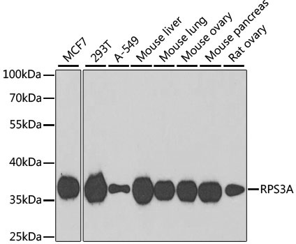
Western blot analysis of various lysates using RPS3A Rabbit pAb (CAB5885) at 1:1000 dilution. Secondary antibody: HRP-conjugated Goat anti-Rabbit IgG (H+L) (CABS014) at 1:10000 dilution. Lysates/proteins: 25μg per lane. Blocking buffer: 3% nonfat dry milk in TBST. Detection: ECL Basic Kit (AbGn00020). Exposure time: 90s.
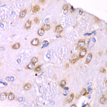
Immunohistochemistry analysis of paraffin-embedded Rat brain using RPS3A Rabbit pAb (CAB5885) at dilution of 1:100 (40x lens). Microwave antigen retrieval performed with 0.01M PBS Buffer (pH 7.2) prior to IHC staining.
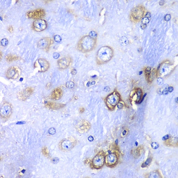
Immunohistochemistry analysis of paraffin-embedded Mouse brain using RPS3A Rabbit pAb (CAB5885) at dilution of 1:100 (40x lens). Microwave antigen retrieval performed with 0.01M PBS Buffer (pH 7.2) prior to IHC staining.
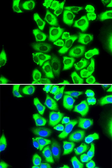
Immunofluorescence analysis of MCF-7 cells using RPS3A Rabbit pAb (CAB5885). Secondary antibody: Cy3-conjugated Goat anti-Rabbit IgG (H+L) (CABS007) at 1:500 dilution. Blue: DAPI for nuclear staining.
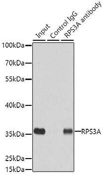
Immunoprecipitation analysis of 200 μg extracts of MCF7 cells using 3 μg RPS3A antibody (CAB5885). Western blot was performed from the immunoprecipitate using RPS3A antibody (CAB5885) at a dilution of 1:500.