Description
SARS-CoV-2 Spike Protein RBD Nanobody [A1]
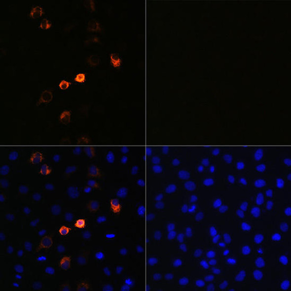
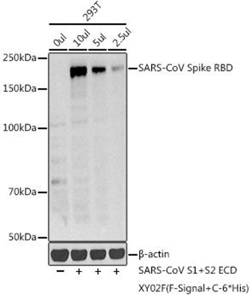
Our SARS-CoV-2 Spike RBD Nanobody is a recombinant monoclonal antibody expressed and synthesized in human embryonic kidney 292 cells (HEK293). This protein is purified from such cells via the use of protein G. A DNA sequence encoding a human IgG1 Fc domain is attached to the end of the sequence encoding the SARS-CoV-2 spike receptor-binding domain (RBD). Protein G allows for the purification of the nanobody by binding to its this IgG1 Fc domain. The small size of this antibody increases the antibody’s ability to bind to relatively inaccessible epitopes on target proteins, ultimately improving its efficacy. This anti-SARS-CoV-2-S1-RBD nanobody functions by targeting the S1 domain of the SARS-CoV-2 spike (S) protein. In doing this it may be used for a range of applications such as ELISA, Colloidal Gold Immunochromatography Assay (GICA), and SARS-CoV-2 neutralization experimentation. Our SARS-CoV-2 Spike RBD Nanobody has been validated with high reactivity towards SARS-CoV-2-S1-RBD by functional ELISA. Experimental analysis has shown it does not exhibit cross-reactivity against RBD domains of other SARS/MERS-related coronaviruses. It is also extremely sensitive, with its sensitivity being confirmed via GICA. The SARS-CoV-2 Spike RBD Nanobody can be used to analyse samples infected with human coronavirus SARS-CoV-2.
Function
Spike glycoprotein comprises two functional subunits responsible for binding to the host cell receptor (S1 subunit) and fusion of the viral and cellular membranes (S2 subunit). For many coronavirus (CoVs), S is cleaved at the boundary between the S1 and S2 subunits, which remain non-covalently bound in the prefusion conformation. The distal S1 subunit comprises the receptor-binding domain(s) and contributes to stabilization of the prefusion state of the membrane-anchored S2 subunit that contains the fusion machinery. S is further cleaved by host proteases at the so-called S2' site located immediately upstream of the fusion peptide in all CoVs. This cleavage has been proposed to activate the protein for membrane fusion via extensive irreversible conformational changes. However, different CoVs use distinct domains within the S1 subunit to recognize a variety of attachment and entry receptors, depending on the viral species. Endemic human coronaviruses OC43 and HKU1 attach via their S domain A to 5-N-acetyl-9-O-acetyl-sialosides found on glycoproteins and glycolipids at the host cell surface to enable entry into susceptible cells. MERS-CoV S uses domain A to recognize non-acetylated sialoside attachment receptors, which likely promote subsequent binding of domain B to the entry receptor, dipeptidyl-peptidase 4. SARS-CoV and several SARS-related coronaviruses (SARSr-CoV) interact directly with angiotensin-converting enzyme 2 (ACE2) via SB to enter target cells.
Information | Description | ||||||||
Uniprot No. | |||||||||
Target Names | S | ||||||||
Antibody Size | 50ul | ||||||||
Alternative Names | Anti-coronavirus spike Antibody; Anti-cov spike Antibody; Anti-ncov RBD Antibody; Anti-ncov S1 Antibody;Anti-ncov spike Antibody; Anti-novel coronavirus RBD Antibody; Anti-novel coronavirus S1 Antibody; Anti-novel coronavirus spike Antibody; Anti-RBD Antibody; Anti-S1 Antibody; Anti-Spike RBD Antibody; E2 Antibody; E2 glycoprotein Antibody; Human coronavirus spike glycoprotein Antibody; S Antibody; SARS-CoV-2 S1 RBD Antibody; S glycoprotein Antibody; Spike glycoprotein Antibody | ||||||||
Species Reactivity | Human Novel Coronavirus (SARS-CoV-2/ 2019-nCoV) | ||||||||
Immunogen | Recombinant Human Novel Coronavirus Spike glycoprotein(S) (319-541aa) | ||||||||
Immunogen Species | Human Novel Coronavirus (SARS-CoV-2/ 2019-nCoV) | ||||||||
Conjugate | Non-conjugated | ||||||||
Clonality | Monoclonal | ||||||||
Isotype | VHH fusion with human IgG1 Fc | ||||||||
Clone No. | A1 | ||||||||
Purification Method | Affinity-chromatography | ||||||||
Concentration | It differs from different batches. Please contact us to confirm it. | ||||||||
Buffer | Preservative: 0.03% Proclin 300 | ||||||||
Form | Liquid | ||||||||
Tested Applications | ELISA, GICA, Neutralising | ||||||||
Recommended Dilution |
| ||||||||
Storage | Upon receipt, store at -20°C or -80°C. Avoid repeated freeze. | ||||||||
Subcellular Location | Virion membrane, Single-pass type I membrane protein, Host endoplasmic reticulum-Golgi intermediate compartment membrane, Single-pass type I membrane protein, Host cell membrane, Single-pass type I membrane protein. | ||||||||
Protein Families | Betacoronaviruses spike protein family |
SARS-CoV-2 Spike glycoprotein Information
Key Information | Description |
Uniprot: | |
Protein: | Spike glycoprotein |
Gene: | S |
Organism: | Severe acute respiratory syndrome coronavirus 2 (2019-nCoV) (SARS-CoV-2) |
Alternative Names: | Spike glycoprotein, S glycoprotein, E2, Peplomer protein |
Spike protein function
Attaches the virion to the cell membrane by interacting with host receptor, initiating the infection (By similarity). Binding to human ACE2 receptor and internalization of the virus into the endosomes of the host cell induces conformational changes in the Spike glycoprotein (PubMed:32142651, PubMed:32075877, PubMed:32155444). Uses also human TMPRSS2 for priming in human lung cells which is an essential step for viral entry (PubMed:32142651). Proteolysis by cathepsin CTSL may unmask the fusion peptide of S2 and activate membranes fusion within endosomes.
Mediates fusion of the virion and cellular membranes by acting as a class I viral fusion protein. Under the current model, the protein has at least three conformational states: pre-fusion native state, pre-hairpin intermediate state, and post-fusion hairpin state. During viral and target cell membrane fusion, the coiled coil regions (heptad repeats) assume a trimer-of-hairpins structure, positioning the fusion peptide in close proximity to the C-terminal region of the ectodomain. The formation of this structure appears to drive apposition and subsequent fusion of viral and target cell membranes.
Acts as a viral fusion peptide which is unmasked following S2 cleavage occurring upon virus endocytosis.
Post-Translational Modification
The cytoplasmic Cys-rich domain is palmitoylated. Spike glycoprotein is digested within host endosomes.
Specific enzymatic cleavages in vivo yield mature proteins. The precurssor is processed into S1 and S2 by host cell furin or another cellular protease to yield the mature S1 and S2 proteins (PubMed:32155444). Additionally, a second cleavage leads to the release of a fusion peptide after viral attachment to host cell receptor (By similarity). The presence of a furin polybasic cleavage site sets SARS-CoV-2 S apart from SARS-CoV S that possesses a monobasic S1/S2 cleavage site processed upon entry of target cells (PubMed:32155444).
Highly decorated by heterogeneous N-linked glycans protruding from the trimer surface.