The Symmetric DiMethyl-Histone H4-R3 Antibody (CAB3159) is a high-quality antibody developed for reliable detection and analysis of target proteins. This antibody, generated in rabbits, specifically targets the symmetric dimethylation of histone H4 at arginine 3 (R3) and is highly reactive with human samples. Validated for use in Western blot applications, this antibody enables the detection and analysis of histone H4 modifications in various cell types.Histone modifications, such as symmetric dimethylation, play a crucial role in gene regulation and chromatin structure, impacting processes like transcription and DNA repair.
This antibody is validated for use in WB, IHC-P, IF/ICC, ELISA, DB applications and has demonstrated reactivity against Human, Mouse, Rat, Other (Wide Range Predicted) samples.
Product Name:
Symmetric DiMethyl-Histone H4-R3 Antibody
SKU:
CAB3159
Size:
20μL, 100μL
Reactivity:
Human, Mouse, Rat, Other (Wide Range Predicted)
Conjugate:
Unconjugated
Immunogen:
Synthetic peptide. This information is considered to be commercially sensitive.
Histones are basic nuclear proteins that are responsible for the nucleosome structure of the chromosomal fiber in eukaryotes. This structure consists of approximately 146 bp of DNA wrapped around a nucleosome, an octamer composed of pairs of each of the four core histones (H2A, H2B, H3, and H4). The chromatin fiber is further compacted through the interaction of a linker histone, H1, with the DNA between the nucleosomes to form higher order chromatin structures. This gene is intronless and encodes a replication-dependent histone that is a member of the histone H4 family. Transcripts from this gene lack polyA tails; instead, they contain a palindromic termination element. This gene is found in a histone cluster on chromosome 1. This gene is one of four histone genes in the cluster that are duplicated; this record represents the centromeric copy.
Purification Method
Affinity purification
Gene ID
8359
RRID
AB_2764952
Buffer Information
Store at -20℃. Avoid freeze / thaw cycles. Buffer: PBS containing 50% glycerol, preserved with proclin300 or sodium azide, pH 7.3.
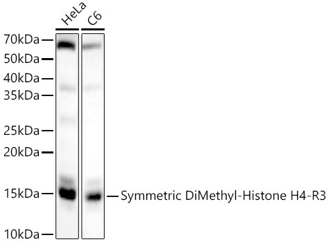
Western blot analysis of various lysates, using Symmetric DiMethyl-Histone H4-R3 Rabbit pAb (CAB3159) at 1:2000 dilution. Secondary antibody: HRP-conjugated Goat anti-Rabbit IgG (H+L) (CABS014) at 1:10000 dilution. Lysates/proteins: 25μg per lane. Blocking buffer: 3% nonfat dry milk in TBST. Detection: ECL Basic Kit (AbGn00020). Exposure time: 90s.
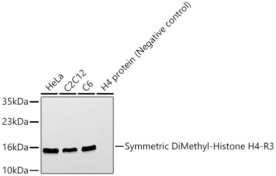
Western blot analysis of various lysates, using Symmetric DiMethyl-Histone H4-R3 Rabbit pAb (CAB3159) at 1:400 dilution. Secondary antibody: HRP-conjugated Goat anti-Rabbit IgG (H+L) (CABS014) at 1:10000 dilution. Lysates/proteins: 25μg per lane. Blocking buffer: 3% nonfat dry milk in TBST. Detection: ECL Basic Kit (AbGn00020). Exposure time: 60s.
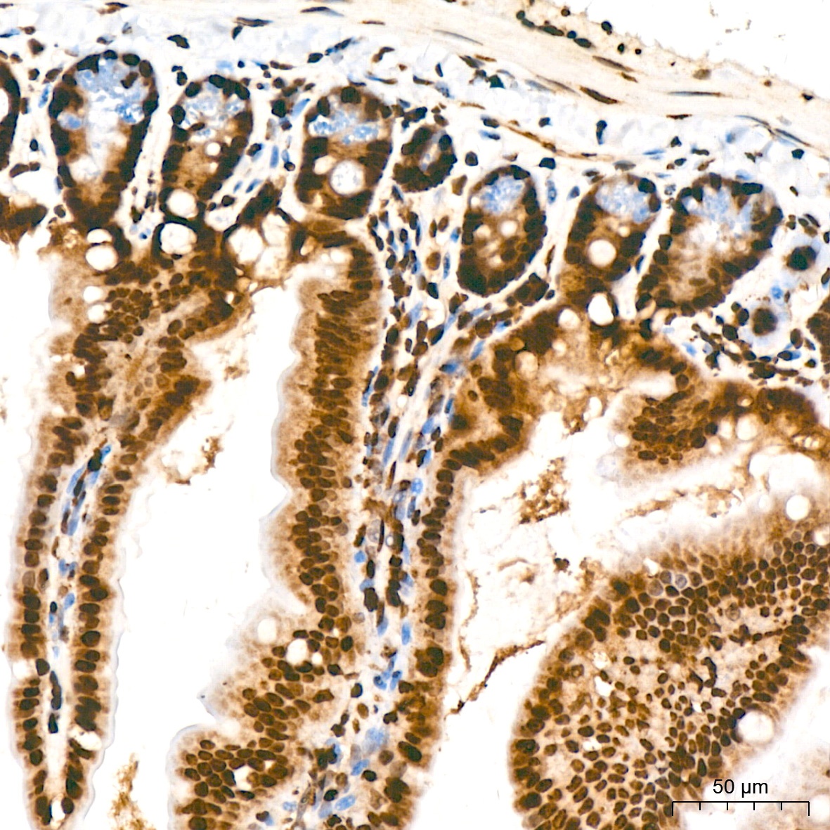
Immunohistochemistry analysis of paraffin-embedded Mouse intestin tissue using Symmetric DiMethyl-Histone H4-R3 Rabbit pAb (CAB3159) at a dilution of 1:100 (40x lens). High pressure antigen retrieval was performed with 0.01 M citrate buffer (pH 6.0) prior to IHC staining.
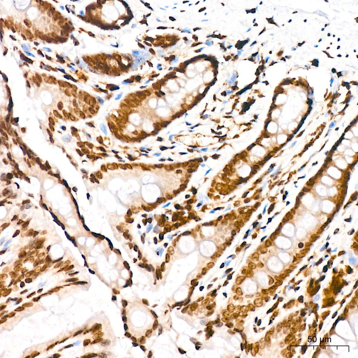
Immunohistochemistry analysis of paraffin-embedded Rat colon tissue using Symmetric DiMethyl-Histone H4-R3 Rabbit pAb (CAB3159) at a dilution of 1:100 (40x lens). High pressure antigen retrieval was performed with 0.01 M citrate buffer (pH 6.0) prior to IHC staining.
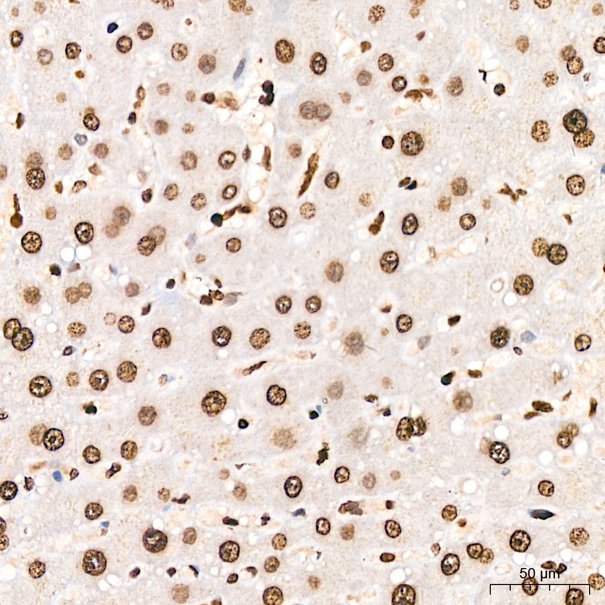
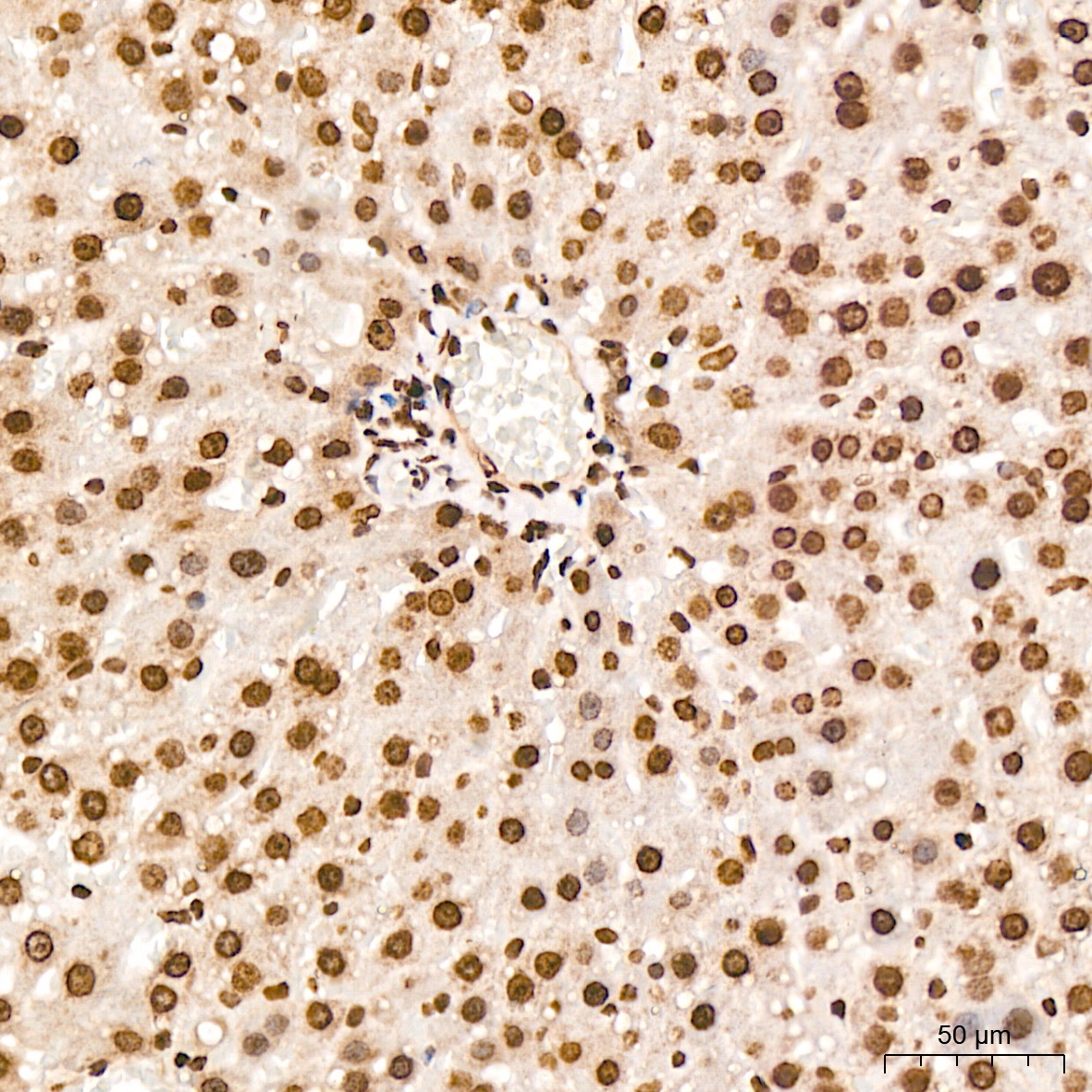
Immunohistochemistry analysis of paraffin-embedded Human liver tissue using Symmetric DiMethyl-Histone H4-R3 Rabbit pAb (CAB3159) at a dilution of 1:100 (40x lens). High pressure antigen retrieval was performed with 0.01 M citrate buffer (pH 6.0) prior to IHC staining.
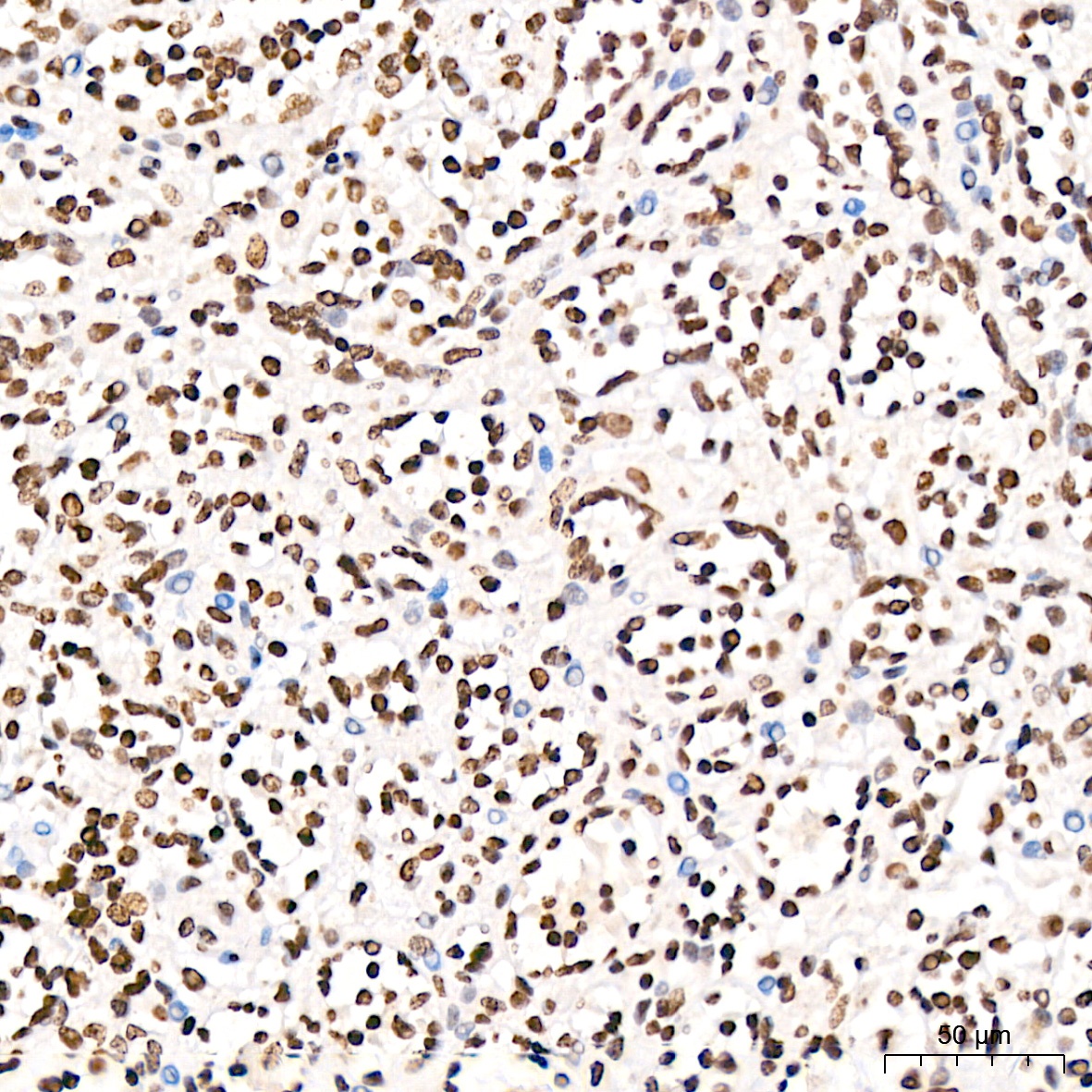
Immunohistochemistry analysis of paraffin-embedded Human cervix cancer tissue using Symmetric DiMethyl-Histone H4-R3 Rabbit pAb (CAB3159) at a dilution of 1:100 (40x lens). High pressure antigen retrieval was performed with 0.01 M citrate buffer (pH 6.0) prior to IHC staining.
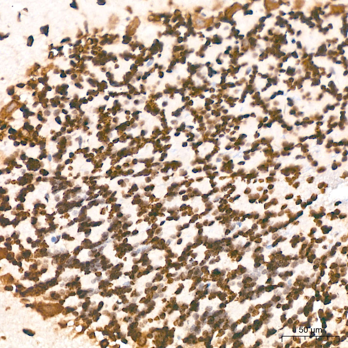
Immunohistochemistry analysis of paraffin-embedded Human spleen tissue using Symmetric DiMethyl-Histone H4-R3 Rabbit pAb (CAB3159) at a dilution of 1:100 (40x lens). High pressure antigen retrieval was performed with 0.01 M citrate buffer (pH 6.0) prior to IHC staining.
Immunohistochemistry analysis of paraffin-embedded Rat brain tissue using Symmetric DiMethyl-Histone H4-R3 Rabbit pAb (CAB3159) at a dilution of 1:100 (40x lens). High pressure antigen retrieval was performed with 0.01 M citrate buffer (pH 6.0) prior to IHC staining.
Immunohistochemistry analysis of paraffin-embedded Rat liver tissue using Symmetric DiMethyl-Histone H4-R3 Rabbit pAb (CAB3159) at a dilution of 1:100 (40x lens). High pressure antigen retrieval was performed with 0.01 M citrate buffer (pH 6.0) prior to IHC staining.
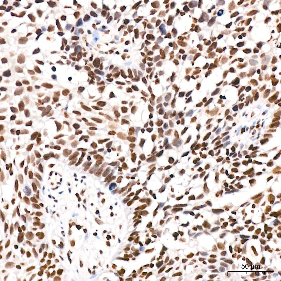
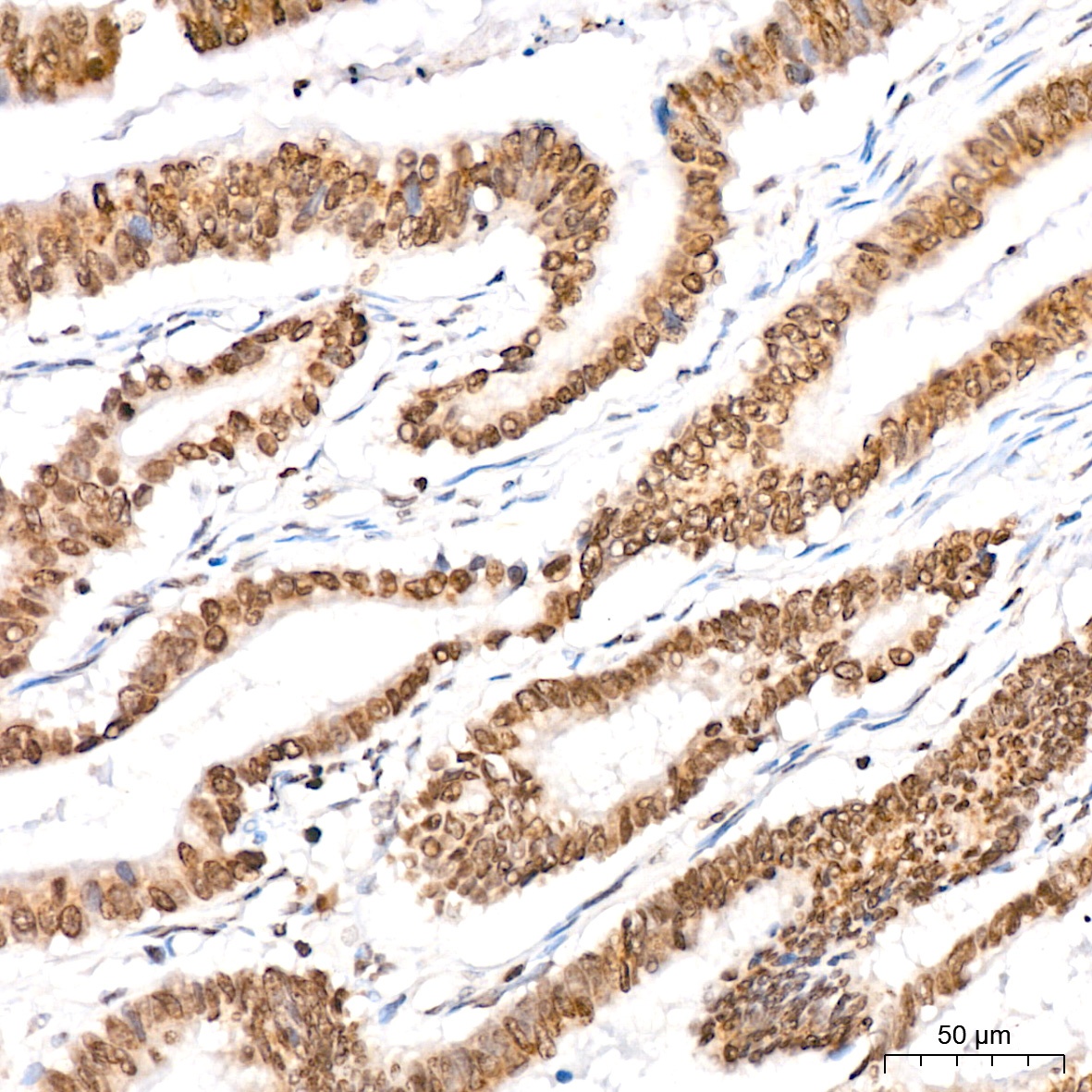
Immunohistochemistry analysis of paraffin-embedded Human colon carcinoma tissue using Symmetric DiMethyl-Histone H4-R3 Rabbit pAb (CAB3159) at a dilution of 1:100 (40x lens). High pressure antigen retrieval was performed with 0.01 M citrate buffer (pH 6.0) prior to IHC staining.
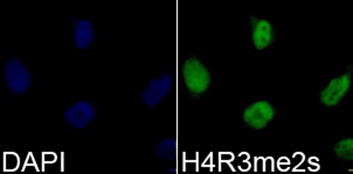
Immunofluorescence analysis of 293T cells using Symmetric DiMethyl-Histone H4-R3 Rabbit pAb (CAB3159). Blue: DAPI for nuclear staining.