The TBX3 Antibody (CAB4144) is a high-quality antibody developed for reliable detection and analysis of target proteins. This antibody is produced in rabbits and exhibits high reactivity with human samples, making it ideal for a variety of research applications.TBX3 is a critical regulator of cell proliferation, differentiation, and migration, with dysregulation implicated in various cancers and developmental disorders. The TBX3 Polyclonal Antibody binds specifically to the TBX3 protein, enabling precise detection and analysis in different cell types and tissues.
This antibody is validated for use in WB, ELISA applications and has demonstrated reactivity against Human, Mouse samples.
Product Name:
TBX3 Antibody
SKU:
CAB4144
Size:
20μL, 100μL
Reactivity:
Human, Mouse
Conjugate:
Unconjugated
Immunogen:
Recombinant protein (or fragment).This information is considered to be commercially sensitive.
Recommended starting concentration is 1 μg/mL. Please optimize the concentration based on your specific assay requirements.
Synonyms:
UMS, XHL, TBX3-ISO, TBX3
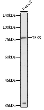
Positive Sample:
HepG2
Cellular Localization:
Nucleus.
Calculated MW:
79kDa
Observed MW:
79kDa
This gene is a member of a phylogenetically conserved family of genes that share a common DNA-binding domain, the T-box. T-box genes encode transcription factors involved in the regulation of developmental processes. This protein is a transcriptional repressor and is thought to play a role in the anterior/posterior axis of the tetrapod forelimb. Mutations in this gene cause ulnar-mammary syndrome, affecting limb, apocrine gland, tooth, hair, and genital development. Alternative splicing of this gene results in three transcript variants encoding different isoforms; however, the full length nature of one variant has not been determined.
Purification Method
Affinity purification
Gene ID
6926
RRID
AB_2765524
Buffer Information
Store at -20℃. Avoid freeze / thaw cycles. Buffer: PBS containing 50% glycerol, preserved with proclin300 or sodium azide, pH 7.3.
Western blot analysis of lysates from HepG2 cells, using TBX3 Rabbit pAb (CAB4144) at 1:1000 dilution. Secondary antibody: HRP-conjugated Goat anti-Rabbit IgG (H+L) (CABS014) at 1:10000 dilution. Lysates/proteins: 25μg per lane. Blocking buffer: 3% nonfat dry milk in TBST. Detection: ECL Basic Kit (AbGn00020). Exposure time: 180s.