The TDP-43/TARDB Monoclonal Antibody (CAB19123) is a high-quality antibody developed for reliable detection and analysis of target proteins. This antibody, produced in rabbits, is highly reactive with human samples and has been validated for use in various applications, including Western blotting and immunofluorescence staining.TDP43 is a key player in RNA metabolism and has been implicated in the pathogenesis of neurodegenerative disorders. By targeting TDP43 with this antibody, researchers can analyze its expression, localization, and function in different cell types and tissues.
This antibody is validated for use in WB, IHC-P, IF/ICC, IP, ELISA, IF-P applications and has demonstrated reactivity against Human, Mouse, Rat samples.
Product Name:
TDP-43/TARDB Monoclonal Antibody
SKU:
CAB19123
Size:
20μL, 100μL
Reactivity:
Human, Mouse, Rat
Clone Number:
ARC0492
Conjugate:
Unconjugated
Immunogen:
Synthetic peptide. This information is considered to be commercially sensitive.
HIV-1, the causative agent of acquired immunodeficiency syndrome (AIDS), contains an RNA genome that produces a chromosomally integrated DNA during the replicative cycle. Activation of HIV-1 gene expression by the transactivator Tat is dependent on an RNA regulatory element (TAR) located downstream of the transcription initiation site. The protein encoded by this gene is a transcriptional repressor that binds to chromosomally integrated TAR DNA and represses HIV-1 transcription. In addition, this protein regulates alternate splicing of the CFTR gene. A similar pseudogene is present on chromosome 20.
Purification Method
Affinity purification
Gene ID
23435
RRID
AB_2862616
Buffer Information
Store at -20℃. Avoid freeze / thaw cycles. Buffer: PBS containing 50% glycerol and 0.05% BSA, preserved with proclin300 or sodium azide, pH 7.3.
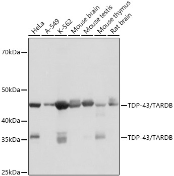
Western blot analysis of various lysates using TDP-43/TARDBP Rabbit mAb (CAB19123) at 1:1000 dilution. Secondary antibody: HRP-conjugated Goat anti-Rabbit IgG (H+L) (CABS014) at 1:10000 dilution. Lysates/proteins: 25μg per lane. Blocking buffer: 3% nonfat dry milk in TBST. Detection: ECL Basic Kit (AbGn00020). Exposure time: 10s.
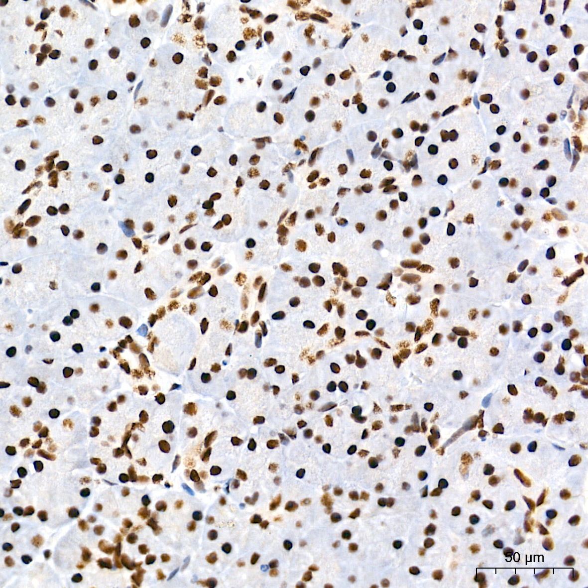
Immunohistochemistry analysis of paraffin-embedded Human pancreas tissue using TDP-43/TARDBP Rabbit mAb (CAB19123) at a dilution of 1:500 (40x lens). High pressure antigen retrieval performed with 0.01M Tris-EDTA Buffer (pH 9.0) prior to IHC staining.
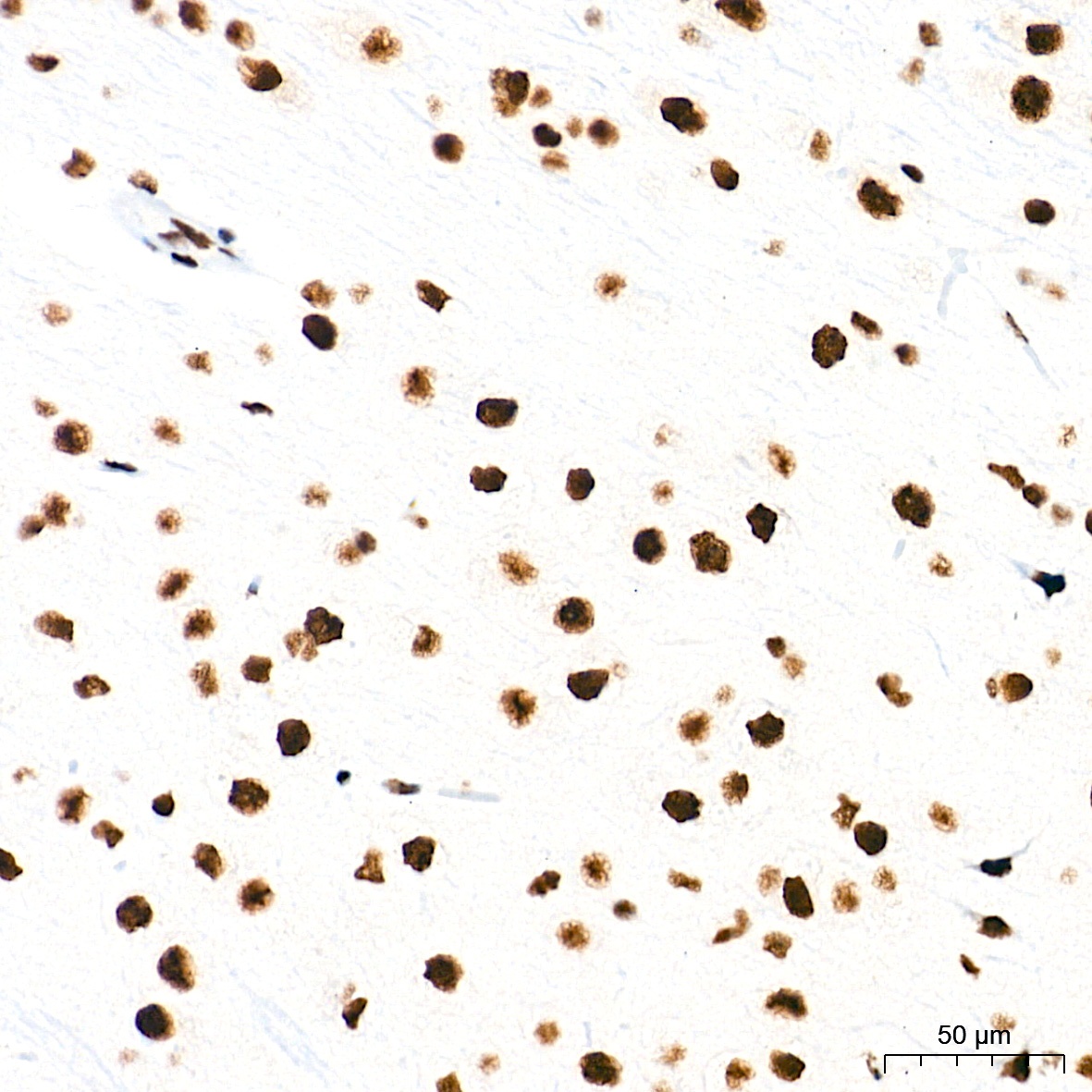
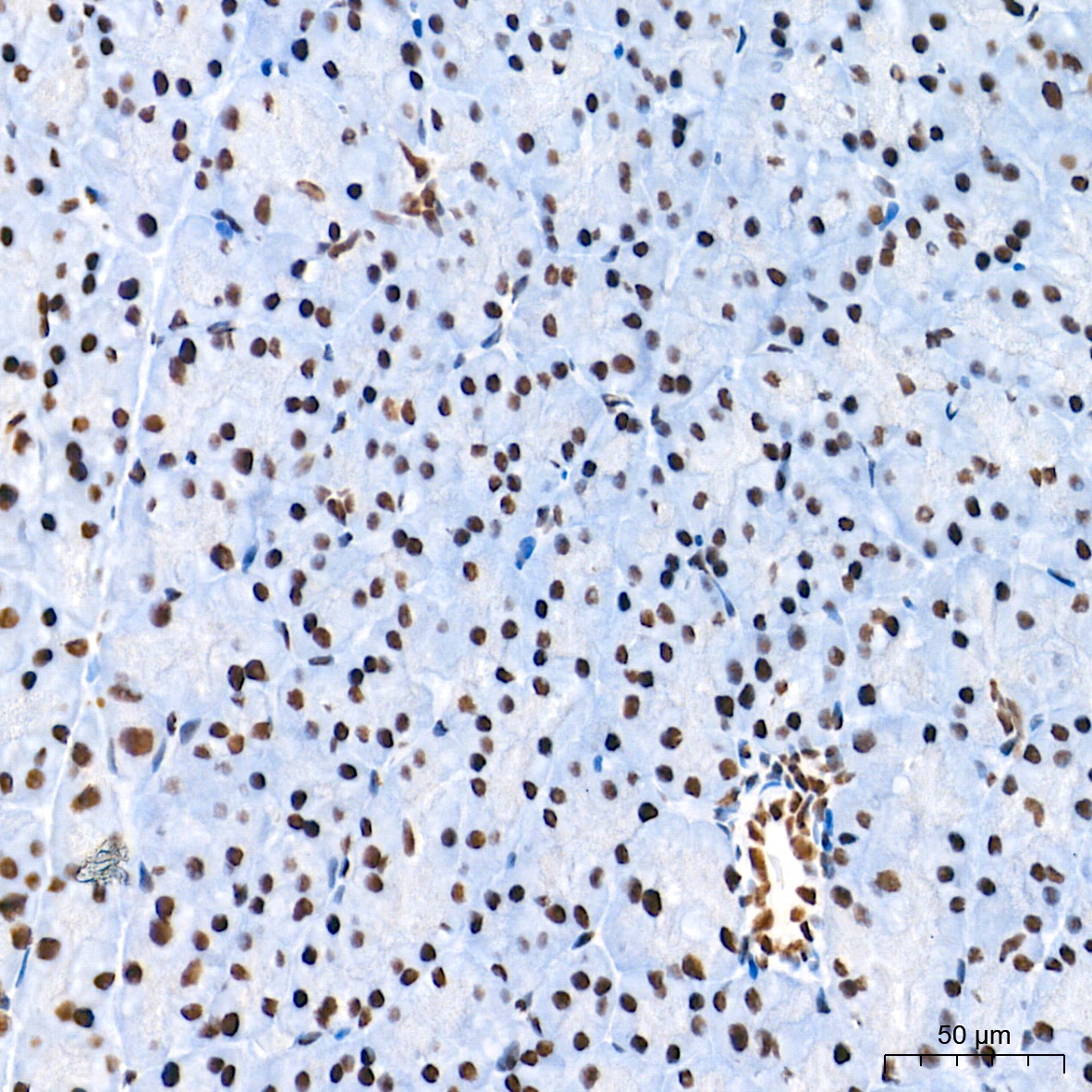
Immunohistochemistry analysis of paraffin-embedded Mouse brain tissue using TDP-43/TARDBP Rabbit mAb (CAB19123) at a dilution of 1:500 (40x lens). High pressure antigen retrieval performed with 0.01M Tris-EDTA Buffer (pH 9.0) prior to IHC staining.
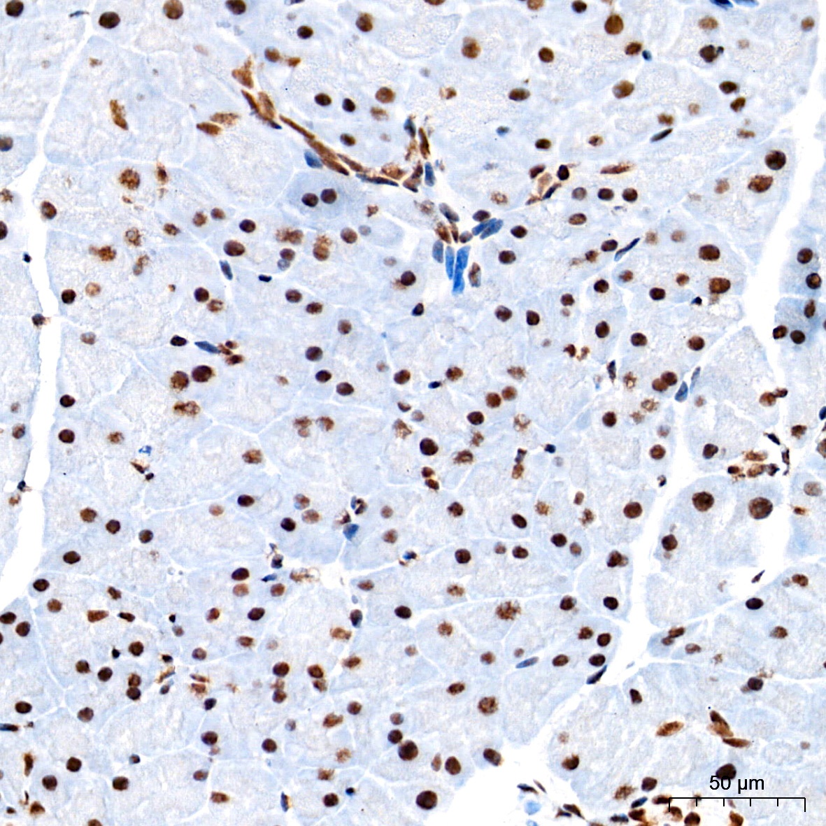
Immunohistochemistry analysis of paraffin-embedded Mouse pancreas tissue using TDP-43/TARDBP Rabbit mAb (CAB19123) at a dilution of 1:500 (40x lens). High pressure antigen retrieval performed with 0.01M Tris-EDTA Buffer (pH 9.0) prior to IHC staining.
Immunohistochemistry analysis of paraffin-embedded Rat pancreas tissue using TDP-43/TARDBP Rabbit mAb (CAB19123) at a dilution of 1:500 (40x lens). High pressure antigen retrieval performed with 0.01M Tris-EDTA Buffer (pH 9.0) prior to IHC staining.
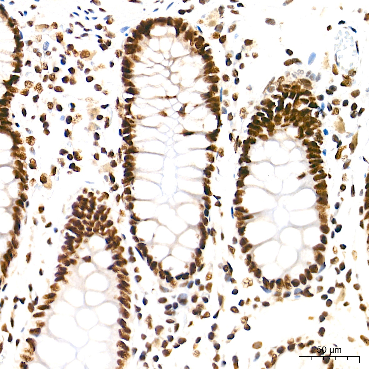
Immunohistochemistry analysis of paraffin-embedded Human colon tissue using TDP-43/TARDBP Rabbit mAb (CAB19123) at a dilution of 1:500 (40x lens). High pressure antigen retrieval performed with 0.01M Tris-EDTA Buffer (pH 9.0) prior to IHC staining.
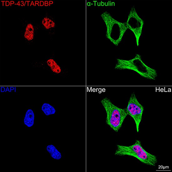
Confocal imaging of HeLa cells using TDP-43/TARDBP Rabbit mAb (CAB19123, dilution 1:200) followed by a further incubation with Cy3 Goat Anti-Rabbit IgG (H+L) (CABS007, dilution 1:500) (Red). The cells were counterstained with α-Tubulin Mouse mAb (AC012, dilution 1:400) followed by incubation with ABflo® 488-conjugated Goat Anti-Mouse IgG (H+L) Ab (CABS076, dilution 1:500) (Green). DAPI was used for nuclear staining (Blue). Objective: 100x.
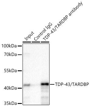
Immunoprecipitation of TDP-43/TARDBP from 500 µg extracts of K-562 cells was performed using 2 µg of TDP-43/TARDBP Rabbit mAb (CAB19123). Rabbit IgG isotype control (AC042) was used to precipitate the Control IgG sample. IP samples were eluted with 1X non-reducing Laemmli Buffer. The Input lane represents 10% of the total input. Western blot analysis of immunoprecipitates was conducted using TDP-43/TARDBP Rabbit mAb (CAB19123) at a dilution of 1:500.