The [KO Validated] TLR4 Antibody (CAB11226) is a high-quality antibody developed for reliable detection and analysis of target proteins. The antibody, produced in rabbits, shows high reactivity with human TLR4 and is validated for use in Western blot and immunohistochemistry applications. By specifically binding to TLR4, this antibody enables precise detection and analysis of TLR4 expression in various cell types, making it indispensable for studies in immunology and infectious disease research.
This antibody is validated for use in WB, IF/ICC, IP, ELISA applications and has demonstrated reactivity against Human, Mouse samples.
Product Name:
[KO Validated] TLR4 Antibody
SKU:
CAB11226
Size:
20μL, 100μL
Reactivity:
Human, Mouse
Conjugate:
Unconjugated
Immunogen:
Recombinant protein (or fragment).This information is considered to be commercially sensitive.
0.5μg-4μg antibody for 200μg-400μg extracts of whole cells
ELISA
Recommended starting concentration is 1 μg/mL. Please optimize the concentration based on your specific assay requirements.
Synonyms:
TOLL, CD284, TLR-4, ARMD10, TLR4
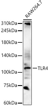
Positive Sample:
RAW264.7
Cellular Localization:
Cell Membrane, Single-Pass Type I Membrane Protein.
Calculated MW:
96kDa
Observed MW:
100-135kDa
The protein encoded by this gene is a member of the Toll-like receptor (TLR) family which plays a fundamental role in pathogen recognition and activation of innate immunity. TLRs are highly conserved from Drosophila to humans and share structural and functional similarities. They recognize pathogen-associated molecular patterns that are expressed on infectious agents, and mediate the production of cytokines necessary for the development of effective immunity. The various TLRs exhibit different patterns of expression. In silico studies have found a particularly strong binding of surface TLR4 with the spike protein of severe acute respiratory syndrome coronavirus 2 (SARS-CoV-2), the causative agent of Coronavirus disease-2019 (COVID-19). This receptor has also been implicated in signal transduction events induced by lipopolysaccharide (LPS) found in most gram-negative bacteria. Mutations in this gene have been associated with differences in LPS responsiveness, and with susceptibility to age-related macular degeneration. Multiple transcript variants encoding different isoforms have been found for this gene.
Purification Method
Affinity purification
Gene ID
7099
RRID
AB_2758466
Buffer Information
Store at -20℃. Avoid freeze / thaw cycles. Buffer: PBS containing 50% glycerol, preserved with proclin300 or sodium azide, pH 7.3.
Western blot analysis of various lysates, using TLR4 Rabbit pAb (CAB11226) at 1:2000 dilution. Secondary antibody: HRP-conjugated Goat anti-Rabbit IgG (H+L) (CABS014) at 1:10000 dilution. Lysates/proteins: 25μg per lane. Blocking buffer: 3% nonfat dry milk in TBST. Detection: ECL Basic Kit (AbGn00020). Exposure time: 180s.
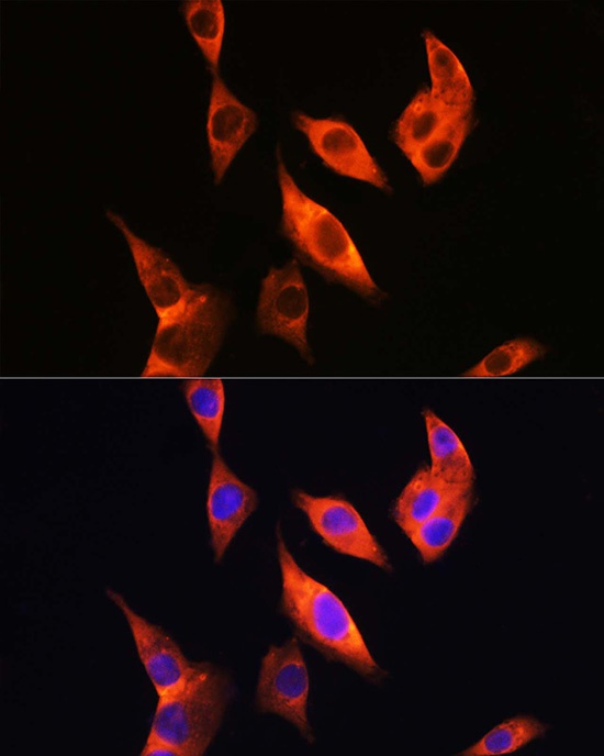
Immunofluorescence analysis of HepG2 cells using TLR4 Rabbit pAb (CAB11226) at dilution of 1:200 (40x lens). Secondary antibody: Cy3-conjugated Goat anti-Rabbit IgG (H+L) (CABS007) at 1:500 dilution. Blue: DAPI for nuclear staining.
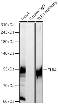
Immunoprecipitation analysis of 300ug extracts of HeLa cells using 3ug TLR4 Rabbit pAb (CAB11226 1:100). Western blot was performed from the immunoprecipitate using TLR4 Rabbit pAb antibody (CAB11226) at a dilition of 1:500.