The TP53I13 Antibody (CAB15924) is a high-quality antibody developed for reliable detection and analysis of target proteins. This antibody, raised in rabbits, is highly reactive with human samples and is validated for use in Western blot applications. It binds to the TP53I13 protein, allowing for accurate detection and analysis in a variety of cell types, making it an excellent tool for studies in molecular biology and cancer research.TP53I13, also known as tumor protein p53-inducible protein 13, is a key player in maintaining genomic stability and promoting cell survival under stress conditions.
This antibody is validated for use in WB, IF/ICC, ELISA applications and has demonstrated reactivity against Mouse, Rat samples.
Product Name:
TP53I13 Antibody
SKU:
CAB15924
Size:
20μL, 100μL
Reactivity:
Mouse, Rat
Conjugate:
Unconjugated
Immunogen:
Recombinant protein (or fragment).This information is considered to be commercially sensitive.
Recommended starting concentration is 1 μg/mL. Please optimize the concentration based on your specific assay requirements.
Synonyms:
DSCP1, TP53I13
Positive Sample:
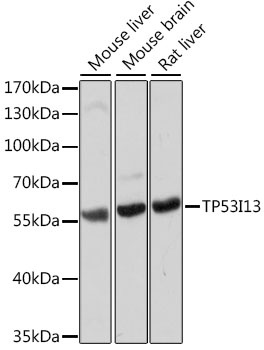
Mouse liver, Mouse brain, Rat liver
Cellular Localization:
Cell Membrane, Cytoplasm, Extracellular Side, Single-Pass Type I Membrane Protein.
Calculated MW:
42kDa
Observed MW:
55kDa
Involved in several processes, including negative regulation of cell cycle; response to UV; and response to xenobiotic stimulus. Located in cytoplasm.
Purification Method
Affinity purification
Gene ID
90313
RRID
AB_2763357
Buffer Information
Store at -20℃. Avoid freeze / thaw cycles. Buffer: PBS with 0.01% thimerosal,50% glycerol,pH7.3.
Western blot analysis of various lysates using TP53I13 Rabbit pAb (CAB15924) at 1:1000 dilution. Secondary antibody: HRP-conjugated Goat anti-Rabbit IgG (H+L) (CABS014) at 1:10000 dilution. Lysates/proteins: 25μg per lane. Blocking buffer: 3% nonfat dry milk in TBST. Detection: ECL Basic Kit (AbGn00020). Exposure time: 10s.
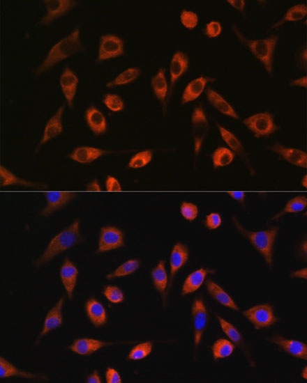
Immunofluorescence analysis of L929 cells using TP53I13 Rabbit pAb (CAB15924) at dilution of 1:100. Secondary antibody: Cy3-conjugated Goat anti-Rabbit IgG (H+L) (CABS007) at 1:500 dilution. Blue: DAPI for nuclear staining.