The GABA transaminase (ABAT) Monoclonal Antibody (CAB9146) is a high-quality antibody developed for reliable detection and analysis of target proteins. This antibody, derived from rabbit monoclonal cells, is specifically designed for use in various applications such as Western blot and immunohistochemistry.ABAT plays a crucial role in GABA metabolism, a neurotransmitter that regulates neuronal excitability in the brain. Dysregulation of ABAT activity has been implicated in neurological disorders such as epilepsy, autism, and schizophrenia.
This antibody is validated for use in WB, IHC-P, ELISA applications and has demonstrated reactivity against Human, Mouse, Rat samples.
Product Name:
GABA transaminase (ABAT) Monoclonal Antibody
SKU:
CAB9146
Size:
20μL, 100μL
Reactivity:
Human, Mouse, Rat
Clone Number:
ARC1450
Conjugate:
Unconjugated
Immunogen:
Synthetic peptide. This information is considered to be commercially sensitive.
Recommended starting concentration is 1 μg/mL. Please optimize the concentration based on your specific assay requirements.
Synonyms:
GABAT, NPD009, GABA-AT, GABA transaminase (ABAT)
Positive Sample:
HepG2, BxPC-3, SH-SY5Y, Mouse kidney, Rat kidney
Cellular Localization:
Mitochondrion Matrix.
Calculated MW:
56kDa
Observed MW:
56kDa
4-aminobutyrate aminotransferase (ABAT) is responsible for catabolism of gamma-aminobutyric acid (GABA), an important, mostly inhibitory neurotransmitter in the central nervous system, into succinic semialdehyde. The active enzyme is a homodimer of 50-kD subunits complexed to pyridoxal-5-phosphate. The protein sequence is over 95% similar to the pig protein. GABA is estimated to be present in nearly one-third of human synapses. ABAT in liver and brain is controlled by 2 codominant alleles with a frequency in a Caucasian population of 0.56 and 0.44. The ABAT deficiency phenotype includes psychomotor retardation, hypotonia, hyperreflexia, lethargy, refractory seizures, and EEG abnormalities. Multiple alternatively spliced transcript variants encoding the same protein isoform have been found for this gene.
Purification Method
Affinity purification
Gene ID
18
RRID
AB_2863670
Buffer Information
Store at -20℃. Avoid freeze / thaw cycles. Buffer: PBS containing 50% glycerol and 0.05% BSA, preserved with proclin300 or sodium azide, pH 7.3.
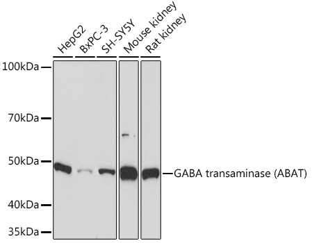
Western blot analysis of various lysates using GABA transaminase (GABA transaminase (ABAT)) Rabbit mAb (CAB9146) at 1:1000 dilution. Secondary antibody: HRP-conjugated Goat anti-Rabbit IgG (H+L) (CABS014) at 1:10000 dilution. Lysates/proteins: 25μg per lane. Blocking buffer: 3% nonfat dry milk in TBST. Detection: ECL Basic Kit (AbGn00020). Exposure time: 10s.
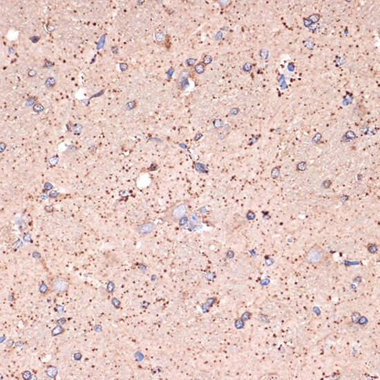
Immunohistochemistry analysis of paraffin-embedded Rat brain using GABA transaminase (GABA transaminase (ABAT)) Rabbit mAb (CAB9146) at dilution of 1:100 (40x lens). Microwave antigen retrieval performed with 0.01M Tris/EDTA Buffer (pH 9.0) prior to IHC staining.
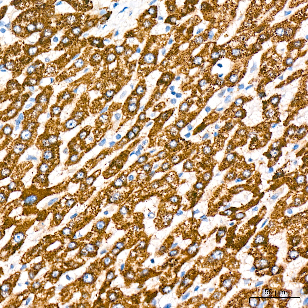
Immunohistochemistry analysis of paraffin-embedded Human liver tissue using GABA transaminase (ABAT) Rabbit mAb (CAB9146) at a dilution of 1:200 (40x lens). High pressure antigen retrieval was performed with 0.01 M citrate buffer (pH 6.0) prior to IHC staining.
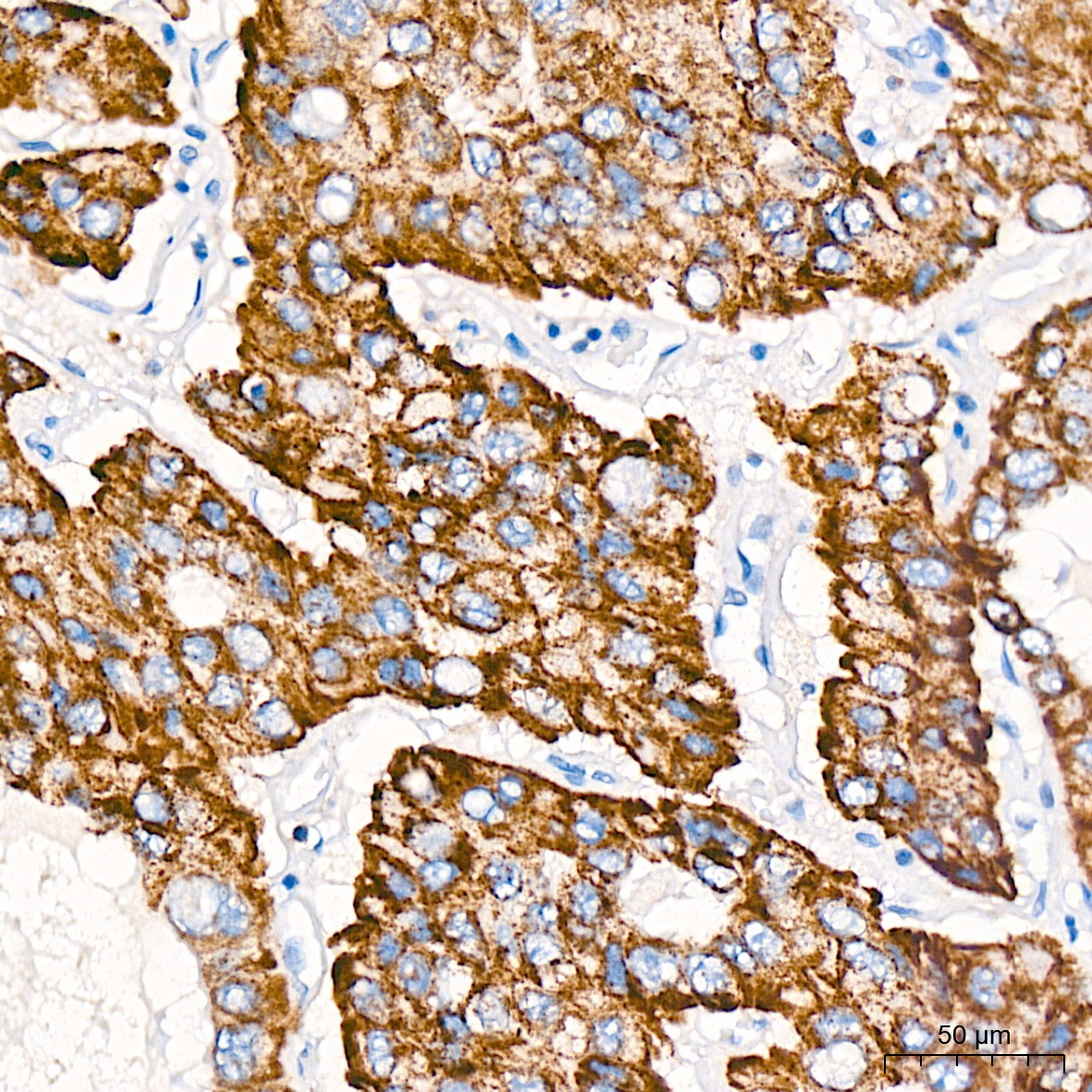
Immunohistochemistry analysis of paraffin-embedded Human liver cancer tissue using GABA transaminase (ABAT) Rabbit mAb (CAB9146) at a dilution of 1:200 (40x lens). High pressure antigen retrieval was performed with 0.01 M citrate buffer (pH 6.0) prior to IHC staining.


![Anti-ABAT [ZN180] Monoclonal Antibody (AGMB06197)](https://cdn11.bigcommerce.com/s-h68l9z2lnx/images/stencil/590x590/products/277478/677449/anti-abat-zn180-monoclonal-antibody-agmb06197__47586.1773032658.jpg?c=2 "Anti-ABAT [ZN180] Monoclonal Antibody (AGMB06197)")
![Anti-ABAT [R04-2E1] Monoclonal Antibody (AGMB01056)](https://cdn11.bigcommerce.com/s-h68l9z2lnx/images/stencil/590x590/products/272345/690879/anti-abat-r04-2e1-monoclonal-antibody-agmb01056__54288.1774501268.jpg?c=2 "Anti-ABAT [R04-2E1] Monoclonal Antibody (AGMB01056)")
![Anti-ABAT [R08-9E3] Monoclonal Antibody (AGMB01055)](https://cdn11.bigcommerce.com/s-h68l9z2lnx/images/stencil/590x590/products/272344/694672/anti-abat-r08-9e3-monoclonal-antibody-agmb01055__74884.1774513179.jpg?c=2 "Anti-ABAT [R08-9E3] Monoclonal Antibody (AGMB01055)")