The CLDN6 Antibody (CAB20465) is a high-quality antibody developed for reliable detection and analysis of target proteins. This polyclonal antibody, produced in rabbits, is highly specific to human samples and is suitable for use in Western blotting, immunofluorescence, and immunohistochemistry applications.CLDN6 is known to be overexpressed in various cancer types, making it a potential therapeutic target for cancer treatment. By using this antibody, researchers can detect and study the expression of CLDN6 in different cell types and tissues, providing valuable insights into its role in cancer progression and metastasis.
This antibody is validated for use in WB, ELISA applications and has demonstrated reactivity against Human, Mouse, Rat samples.
Product Name:
CLDN6 Antibody
SKU:
CAB20465
Size:
20μL, 100μL
Reactivity:
Human, Mouse, Rat
Conjugate:
Unconjugated
Immunogen:
Synthetic peptide. This information is considered to be commercially sensitive.
Tight junctions represent one mode of cell-to-cell adhesion in epithelial or endothelial cell sheets, forming continuous seals around cells and serving as a physical barrier to prevent solutes and water from passing freely through the paracellular space. These junctions are comprised of sets of continuous networking strands in the outwardly facing cytoplasmic leaflet, with complementary grooves in the inwardly facing extracytoplasmic leaflet. This gene encodes a component of tight junction strands, which is a member of the claudin family. The protein is an integral membrane protein and is one of the entry cofactors for hepatitis C virus. The gene methylation may be involved in esophageal tumorigenesis. This gene is adjacent to another family member CLDN9 on chromosome 16.
Purification Method
Affinity purification
Gene ID
9074
Buffer Information
Store at -20℃. Avoid freeze / thaw cycles. Buffer: PBS containing 50% glycerol, preserved with proclin300 or sodium azide, pH 7.3.
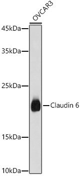
Western blot analysis of lysates from OVCAR3 cells, using Claudin 6 Rabbit pAb (CAB20465) at 1:1000 dilution. Secondary antibody: HRP-conjugated Goat anti-Rabbit IgG (H+L) (CABS014) at 1:10000 dilution. Lysates/proteins: 25μg per lane. Blocking buffer: 3% nonfat dry milk in TBST. Detection: ECL Basic Kit (AbGn00020). Exposure time: 10s.
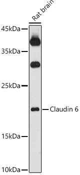
Western blot analysis of lysates from Rat brain, using Claudin 6 Rabbit pAb (CAB20465) at 1:1000 dilution. Secondary antibody: HRP-conjugated Goat anti-Rabbit IgG (H+L) (CABS014) at 1:10000 dilution. Lysates/proteins: 25μg per lane. Blocking buffer: 3% nonfat dry milk in TBST. Detection: ECL Basic Kit (AbGn00020). Exposure time: 180s.