The MT-ND1 Monoclonal Antibody (CAB9743) is a high-quality antibody developed for reliable detection and analysis of target proteins. This antibody, produced in rabbits, has been shown to be highly reactive with human samples and is validated for use in applications such as Western blot and immunohistochemistry.MT-ND1 is a subunit of the mitochondrial respiratory chain complex I, which plays a crucial role in the process of oxidative phosphorylation and ATP production. Dysfunctions in this complex have been associated with various mitochondrial diseases, making MT-ND1 a key target for research in areas such as mitochondrial biology, neurodegenerative diseases, and metabolic disorders.
This antibody is validated for use in WB, ELISA applications and has demonstrated reactivity against Mouse, Rat samples.
Product Name:
MT-ND1 Monoclonal Antibody
SKU:
CAB9743
Size:
20μL, 100μL
Reactivity:
Mouse, Rat
Clone Number:
ARC1728
Conjugate:
Unconjugated
Immunogen:
Synthetic peptide. This information is considered to be commercially sensitive.
Sequence:
Email for sequence
Tested Applications:
WBELISA
Recommended Dilution:
WB
1:500 - 1:1000
ELISA
Recommended starting concentration is 1 μg/mL. Please optimize the concentration based on your specific assay requirements.
Synonyms:
MTND1, ND1, MT-ND1
Positive Sample:
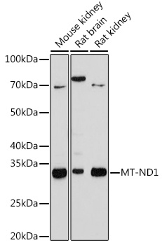
Mouse kidney, Rat brain, Rat kidney
Cellular Localization:
Mitochondrial Inner Membrane, Mitochondrial Membrane, Mitochondrial Respiratory Chain Complex I.
Calculated MW:
36kDa
Observed MW:
35kDa
Enables NADH dehydrogenase (ubiquinone) activity. Involved in mitochondrial electron transport, NADH to ubiquinone and mitochondrial respiratory chain complex I assembly. Located in mitochondrial membrane. Part of mitochondrial respiratory chain complex I. Implicated in several diseases, including MELAS syndrome; neurodegenerative disease (multiple); optic nerve disease (multiple); toxic shock syndrome; and type 2 diabetes mellitus. Biomarker of Alzheimer's disease; Parkinson's disease; and multiple sclerosis.
Purification Method
Affinity purification
Gene ID
4535
Buffer Information
Store at -20℃. Avoid freeze / thaw cycles. Buffer: PBS containing 50% glycerol and 0.05% BSA, preserved with proclin300 or sodium azide, pH 7.3.
Western blot analysis of various lysates using MT-ND1 Rabbit mAb (CAB9743) at 1:1000 dilution. Secondary antibody: HRP-conjugated Goat anti-Rabbit IgG (H+L) (CABS014) at 1:10000 dilution. Lysates/proteins: 25μg per lane. Blocking buffer: 3% nonfat dry milk in TBST. Detection: ECL Basic Kit (AbGn00020). Exposure time: 180s.


![Anti-MT ND1 [R08-8Q9] Monoclonal Antibody (AGMB01773)](https://cdn11.bigcommerce.com/s-h68l9z2lnx/images/stencil/590x590/products/273062/679424/anti-mt-nd1-r08-8q9-monoclonal-antibody-agmb01773__67404.1773038896.jpg?c=2 "Anti-MT ND1 [R08-8Q9] Monoclonal Antibody (AGMB01773)")


