The SARS-CoV-2 Spike Antibody (CAB20137) is a high-quality antibody developed for reliable detection and analysis of target proteins. This antibody, developed and validated by Assay Genie, specifically targets the spike protein of the SARS-CoV-2 virus. The spike protein is essential for viral entry into host cells, making it a prime target for vaccine development and therapeutic strategies. The Anti-SARS-CoV-2 Spike Antibody is highly specific and effective in detecting the spike protein in various sample types, including patient samples and laboratory cultures.
This antibody is validated for use in WB, IF/ICC, IP, ELISA, DB applications and has demonstrated reactivity against SARS-CoV-2 samples.
Product Name:
SARS-CoV-2 Spike Antibody
SKU:
CAB20137
Size:
20μL, 100μL
Reactivity:
SARS-CoV-2
Immunogen:
Synthetic peptide. This information is considered to be commercially sensitive.
0.5μg-4μg antibody for 200μg-400μg extracts of whole cells
ELISA
Recommended starting concentration is 1 μg/mL. Please optimize the concentration based on your specific assay requirements.
Synonyms:
spike glycoprotein, SARS-CoV-2 Spike
Positive Sample:
293T
Calculated MW:
141kDa
Observed MW:
110kDa/180kDa
Severe acute respiratory syndrome coronavirus 2 (SARS-CoV-2) is an enveloped, positive-sense, single-stranded RNA virus that causes coronavirus disease 2019 (COVID-19). Virus particles include the RNA genetic material and structural proteins needed for invasion of host cells. Once inside the cell the infecting RNA is used to encode structural proteins that make up virus particles, nonstructural proteins that direct virus assembly, transcription, replication and host control and accessory proteins whose function has not been determined.~ The structural proteins of SARS-CoV-2 include the envelope protein (E), spike or surface glycoprotein (S), membrane protein (M) and the nucleocapsid protein (N). The spike glycoprotein is found on the outside of the virus particle and gives coronavirus viruses their crown-like appearance. This glycoprotein mediates attachment of the virus particle and entry into the host cell. S protein is an important target for vaccine development, antibody therapies and diagnostic antigen-based tests.
Purification Method
Affinity purification
Gene ID
43740568
RRID
AB_2862929
Buffer Information
Store at -20℃. Avoid freeze / thaw cycles. Buffer: PBS containing 50% glycerol, preserved with proclin300 or sodium azide, pH 7.3.
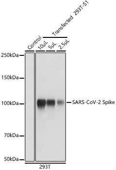
Western blot analysis of extracts of 293T and transfected 293T-S1(His-tag), using SARS-CoV-2 Spike Rabbit pAb (CAB20137) at 1:1000 dilution. Secondary antibody: HRP-conjugated Goat anti-Rabbit IgG (H+L) (CABS014) at 1:10000 dilution. Lysates/proteins: 25μg per lane. Blocking buffer: 3% nonfat dry milk in TBST. Detection: ECL Basic Kit (AbGn00020). Exposure time: 1s.
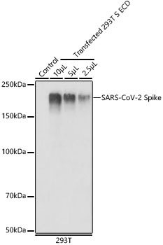
Western blot analysis of extracts of 293T and transfected 293T-S ECD(His-tag), using SARS-CoV-2 Spike Rabbit pAb (CAB20137) at 1:1000 dilution. Lysates/proteins: 25μg per lane. Blocking buffer: 3% nonfat dry milk in TBST. Detection: ECL Basic Kit (AbGn00020). Exposure time: 1s.
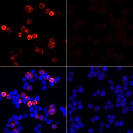
Immunofluorescence analysis of 293T cells transfected with SARS-CoV-2 Spike protein and untreated 293T cells use SARS-CoV-2 Spike Rabbit pAb (CAB20137) at dilution of 1:100 (40x lens). Secondary antibody: Cy3-conjugated Goat anti-Rabbit IgG (H+L) (CABS007) at 1:500 dilution. Blue: DAPI for nuclear staining.
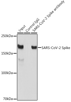
Immunoprecipitation analysis of 300 μg extracts of 293T cells using 3 μg SARS-CoV-2 Spike antibody (CAB20137). Western blot was performed from the immunoprecipitate using SARS-CoV-2 Spike antibody (CAB20137) at a dilution of 1:1000.