
A Comprehensive Overview of Cell Death
Cell death is an essential aspect of cellular biology that plays a crucial role in development, homeostasis, and disease. In this blog, we try to focus on mechanisms and various types of cell death, shedding light on the scientific underpinnings of apoptosis, necrosis, and regulated necrosis variants.
Table of Contents
Jump to a section:
Apoptosis
Apoptosis, a form of cellular demise, ensues when a cell's apoptotic program is set into motion. This program is essentially a sequence of encoded directives stored within the cell's DNA. Upon initiation of apoptosis, the cell undergoes a cascade of transformations that culminate in its demise. Once this process is set in motion, it becomes irreversible, rendering any rescue attempts futile. Apoptosis, a natural occurrence, is a constant phenomenon within our bodies. It serves to eliminate aged, impaired, or redundant cells. Moreover, apoptosis holds pivotal roles in developmental processes and immune responses.
How to Measure Apoptosis
Phenotypic alterations and the presence of specific proteins set apoptotic cells apart from viable ones. Multiple techniques are available to assess and quantify apoptotic occurrences within a cell population. These methodologies encompass
| Assay | Description | Sample Type | Read-Out |
|
In the assay, a specific substrate (N-Ac-DEVD-AFC) is cleaved by active caspase-3, forming a highly fluorescent product |
Cell and Tissue Lysate |
Fluorometric |
|
|
Depolarization and subsequent decrease of the mitochondrial membrane potential is a hallmark feature of apoptotic cells |
Cell Sampes |
Fluoromteric |
|
|
DNA fragmentation which is a hallmark of apoptosis |
Paraffin section, frozen section, cell slide |
Colorimetric/Fluorometric |
|
|
Detects changes that occur to the lipid bilayer early in apoptosis |
Cell samples |
Fluorometric |
|
|
Decreased levels of ATP and increased levels of ADP represent apoptotic cells |
Cell samples |
Fluorometric |
Necrosis
Necrosis represents a pathological sequence characterized by the demise of cells or tissues within a living organism. This phenomenon arises due to an array of factors, including injury, infection, insufficient blood supply (ischemia), toxins, or underlying diseases. Unlike programmed cell death (apoptosis), which adheres to controlled and organized patterns, necrosis tends to be erratic and unregulated. The repercussions of necrosis span from localized tissue harm to broader organ malfunction, contingent upon the scale and location of the impacted cells or tissues. Grasping the origins, manifestations, and potential complexities of necrosis holds paramount importance in the diagnosis and treatment of diverse medical conditions. Furthermore, exploring preventive strategies can play a pivotal role in reducing the susceptibility to necrosis and safeguarding overall health and well-being.
Fig 1: Differences between Apoptosis and Necrosis
How to Measure Necrosis
Multiple techniques are available to assess and quantify necrotic occurrences within a cell population. These methodologies encompass:
Necrosis Related ELISA Kits
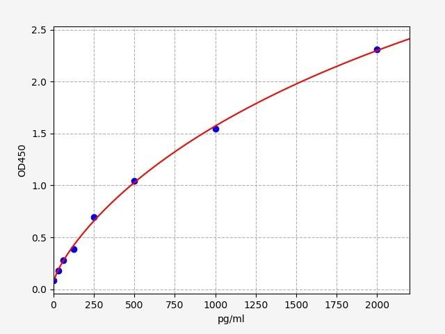
| Human Tumor Necrosis Factor(TNF) ELISA Kit | |
|---|---|
| Size | 96 Assays |
| Range | 15.6-1000 pg/mL |
| ELISA Type | Sandwich |
The Human Tumor Necrosis Factor (TNF) ELISA Kit is a specialized tool designed for the quantitative detection of TNF in human biological samples. This ELISA kit offers a sensitive and accurate means to measure TNF levels, enabling researchers and clinicians to gain insights into the inflammatory response and various disease conditions where TNF plays a pivotal role. With its precise measurement capabilities, the TNF ELISA Kit contributes to a deeper understanding of immune system dynamics and potential therapeutic interventions.
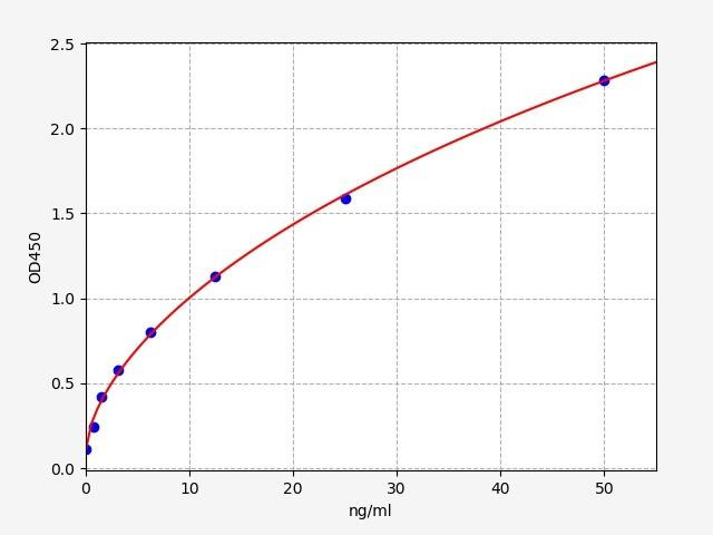
| Human LDHA / L-lactate dehydrogenase A chain ELISA Kit | |
|---|---|
| Sensitivity | 0.469ng/ml |
| Range | 0.781-50ng/ml |
| ELISA Type | Sandwich |
LDHA / L-lactate dehydrogenase A chain is found predominantly in muscle tissue, and catalyzes the conversion of L-lactate and NAD to pyruvate and NADH in the final step of anaerobic glycolysis. Mutations in LDHA / L-lactate dehydrogenase A chain have been linked to Fanconi-Bickel syndrome and exertional myoglobinuria. The Assay Genie Human LDHA / L-lactate dehydrogenase A chain ELISA Kit is a highly sensitive assay for the quantitative measurement of LDHA / L-lactate dehydrogenase A chain in serum, blood, plasma, cell culture supernatant and tissue samples.
High mobility group protein B1 (HMGB1) is a member of the High Mobility Group-box superfamily. HMGB1 has also been shown to act as a danger associated molecular pattern (DAMP) molecule, which enhances the immune response during tissue injury. HMGB1 is believed to play a role in the development of several chronic inflammatory and autoimmune diseases, as well as cancer. The Assay Genie Human HMGB1 ELISA kit is a highly senstive assay for the quantitative measurement of HMGB1 in serum, plasma, cell and tissue lysates.
Types of Regulated Necrosis
Regulated necrosis encompasses various distinct forms of cell death, each characterized by specific molecular mechanisms and physiological triggers. Some prominent types of regulated necrosis include:
- Necroptosis: Necroptosis is a form of regulated necrosis that involves a programmed cell death process with distinct molecular components and triggers. Unlike apoptosis, which is characterized by controlled cellular dismantling, necroptosis results in cell rupture and release of cellular contents, which can trigger an inflammatory response. It is often referred to as "programmed necrosis" because it shares features of both apoptosis and necrosis. Necroptosis serves as a backup mechanism when apoptotic pathways are compromised, such as when caspases are inhibited. It has been implicated in various physiological and pathological conditions, including inflammation, immunity, and tissue repair. Dysregulation of necroptosis has been associated with diseases like neurodegenerative disorders, viral infections, and inflammatory diseases.
- Pyroptosis: Pyroptosis is another form of regulated necrosis that occurs primarily in immune cells as a response to microbial infections. It is characterized by cell swelling, membrane rupture, and the release of pro-inflammatory contents. Pyroptosis is mediated by inflammasomes, which are multiprotein complexes that activate inflammatory caspases, particularly caspase-1 or caspase-11, depending on the cell type and species. Pyroptosis serves as a defense mechanism against intracellular pathogens by releasing antimicrobial peptides and initiating an inflammatory response that attracts immune cells. However, excessive or dysregulated pyroptosis can contribute to tissue damage and inflammatory diseases. Research into pyroptosis has implications for understanding infectious diseases, autoimmunity, and potential therapeutic interventions.
How to Measure Necroptosis
Necroptosis Related ELISA Kits
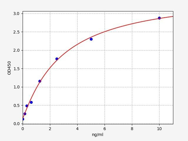
| Human RIPK1 (Receptor interacting serine/threonine kinase 1) ELISA Kit | |
|---|---|
| Sensitivity | 0.094ng/ml |
| Range | 0.156-10ng/ml |
| ELISA Type | Sandwich |
RIPK1 (Receptor interacting serine/threonine kinase 1) encodes a member of the receptor-interacting protein (RIP) family of serine/threonine protein kinases. RIPK1 (Receptor interacting serine/threonine kinase 1) plays a role in inflammation and cell death in response to tissue damage, pathogen recognition, and as part of developmental regulation. Diseases associated with RIPK1 (Receptor interacting serine/threonine kinase 1) include Immunodeficiency, autoinflammation with episodic fever and lymphadenopathy.
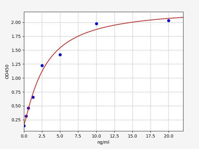
| Human MLKL (mixed lineage kinase domain-like) ELISA Kit | |
|---|---|
| Sensitivity | 0.188ng/ml |
| Range | 0.313-20ng/ml |
| ELISA Type | Sandwich |
MLKL (mixed lineage kinase domain-like) belongs to the protein kinase superfamily. MLKL (mixed lineage kinase domain-like) plays a critical role in tumor necrosis factor (TNF)-induced necroptosis, a programmed cell death process, via interaction with receptor-interacting protein 3 (RIP3). High levels of MLKL (mixed lineage kinase domain-like) are associated with inflammatory bowel disease and diabetes. The Assay Genie Human MLKL (mixed lineage kinase domain-like) ELISA is a highly sensitive assay for the quantitative measurement of MLKL (mixed lineage kinase domain-like) in serum, blood, plasma, cell culture supernatant and tissue samples.
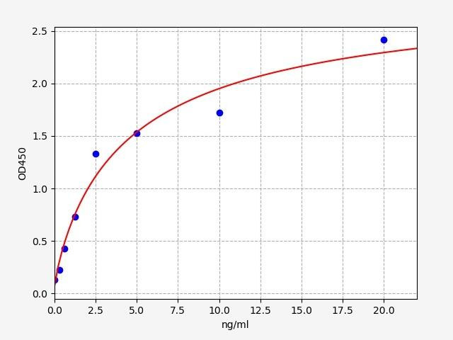
| Human RIPK3 (Receptor-interacting serine/threonine-protein kinase 3) ELISA Kit | |
|---|---|
| Sensitivity | 0.188ng/ml |
| Range | 0.313-20ng/ml |
| ELISA Type | Sandwich |
RIPK3 (Receptor-interacting serine/threonine-protein kinase 3) is predominantly localized to the cytoplasm, and can undergo nucleocytoplasmic shuttling dependent on novel nuclear localization and export signals. RIPK3 (Receptor-interacting serine/threonine-protein kinase 3) is a component of the tumor necrosis factor (TNF) receptor-I signaling complex, and can induce apoptosis by weakly activating the NF-kappaB transcription factor. Diseases associated with RIPK3 (Receptor-interacting serine/threonine-protein kinase 3) include herpes simplex virus infections and caspase 8 deficiency
How to Measure Pyroptosis
| Marker | Description |
|
Gasdermin cleavage |
Cleavage of gasdermin-D occurs during pyroptosis lead to pore formation |
|
Pyroptosis is characterized by the formation of the inflammasome; a marker for the inflammasome is NLRP3 |
|
|
Cleavage of caspase-1 is a marker for its activity. Activated caspase-1 cleaves IL-1β and gasdermin D |
In conclusion, this review focuses on the orchestrated process of apoptosis and necrosis, including regulated forms, a deeper understanding of these phenomena is crucial in deciphering their roles across various biological scenarios. By exploring measurement techniques, we open the door to unraveling the complexities of cell death and its far-reaching implications, ultimately contributing to advancements in both fundamental research and potential therapeutic strategies.
Written by Pragna Krishnapur
Pragna Krishnapur completed her bachelor degree in Biotechnology Engineering in Visvesvaraya Technological University before completing her masters in Biotechnology at University College Dublin.
Recent Posts
-
ELISA vs Western Blot: Which Technique Should You Choose?
Written by Seán Mac Fhearraigh, PhD • Updated: 19 May 2026 • ~9 min read Quick Answer ELISA …20th May 2026 -
Types of ELISA: Direct, Indirect, Sandwich & Competitive Compared
Written by Seán Mac Fhearraigh, PhD • Updated: 19 May 2026 • ~9 min read Quick Answer The fi …20th May 2026 -
Sandwich ELISA: Step-by-Step Protocol & Troubleshooting
Written by Seán Mac Fhearraigh, PhD • Updated: 19 May 2026 • ~ …20th May 2026