
Granulocytes: Neutrophils, Eosinophils, Basophils & Mast Cells
Granulocytes: Neutrophils, Eosinophils and Basophils
Granulocytes are the leukocytes with cytoplasmic granules and lobed nuclei — neutrophils, eosinophils and basophils. They are the fastest-responding arm of cellular immunity, and between them they handle bacterial infection, parasites and allergic inflammation. This guide covers each type, their granule contents and surface markers, how they develop and are recruited, and what immature granulocytes on a blood count actually mean.
Browse granulocyte reagents →Key takeaways
- There are three granulocytes — neutrophils, eosinophils and basophils. They are also called polymorphonuclear leukocytes because of their multilobed nuclei.
- Mast cells are not granulocytes in the standard classification. They are granulated tissue-resident cells of a separate lineage with a single round nucleus, covered here because they pair functionally with basophils.
- Neutrophils dominate numerically and act by phagocytosis, reactive oxygen species and extracellular traps; the NADPH oxidase step is what fails in chronic granulomatous disease.
- Eosinophils target helminths and drive the late phase of allergic inflammation through major basic protein and eosinophil cationic protein.
- Basophils are the rarest leukocyte and, like mast cells, carry high-affinity IgE receptor and release histamine.
- Recruitment follows a fixed sequence — selectin-mediated rolling, integrin-mediated firm adhesion, then chemokine-directed transmigration.
- “Immature granulocytes” on a modern analyser is not the same as the band count: the IG parameter counts promyelocytes, myelocytes and metamyelocytes, and excludes bands.
Contents
Granulocyte reagents
Surface markers for identifying each population by flow cytometry, plus the granule proteins each cell type releases.
![Purified Anti-Human CD66b Antibody [G10F5]](https://cdn11.bigcommerce.com/s-h68l9z2lnx/products/271210/images/691630/purified-anti-human-cd66b-antibody-g10f5-agel5206__75573.1774503575.386.513.jpg?c=2)
Purified Anti-Human CD66b Antibody [G10F5]
A granulocyte-restricted activation marker, widely used to identify and gate neutrophils.
View product →![PerCP/Cyanine5.5 Anti-Human CD16 Antibody [3G8]](https://cdn11.bigcommerce.com/s-h68l9z2lnx/products/229826/images/602870/percpcyanine5.5-anti-human-cd16-antibody-3g8__30145.1706286065.386.513.jpg?c=2)
PerCP/Cyanine5.5 Anti-Human CD16 Antibody [3G8]
High on mature neutrophils and absent from eosinophils — the standard way to separate the two.
View product →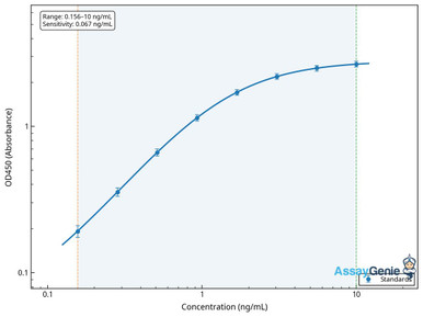
Human Myeloperoxidase (MPO) ELISA Kit
The defining azurophilic granule enzyme of neutrophils and a marker of degranulation.
View product →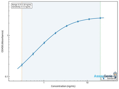
Human Eosinophil Cationic Protein (RNASE3) ELISA Kit
An eosinophil-specific granule protein used as a marker of eosinophil activation in allergic disease.
View product →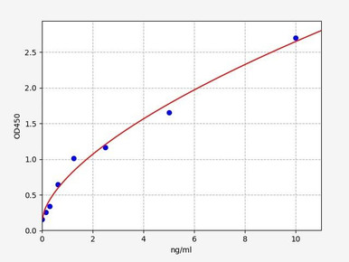
Human Tryptase ELISA Kit
The principal mast cell protease; serum tryptase is the standard marker of mast cell activation.
View product →![PE Anti-Human CD117 Antibody [104D2]](https://cdn11.bigcommerce.com/s-h68l9z2lnx/products/22309/images/606475/pe-anti-human-cd117-antibody-104d2-agel1778__71407.1707498173.386.513.jpg?c=2)
PE Anti-Human CD117 Antibody [104D2]
KIT, the stem cell factor receptor — required for mast cell development and used to identify them.
View product →What granulocytes are
Granulocytes are white blood cells characterised by prominent cytoplasmic granules — membrane-bound vesicles packed with enzymes, antimicrobial peptides and inflammatory mediators. They are also called polymorphonuclear leukocytes, because their nuclei are divided into two or more connected lobes rather than being round.
That nuclear shape is the feature that separates them from the mononuclear leukocytes — lymphocytes and monocytes — and it is why a density gradient preparation of mononuclear cells specifically excludes them.
They are the fast arm of cellular immunity. Neutrophils in particular reach a site of infection within minutes to hours, long before an adaptive response can be mounted.
Three granulocytes — and where mast cells fit
Standard classification recognises three granulocytes, distinguished historically by how their granules take up histological stains.
| Cell | Granule staining | Share of circulating leukocytes |
|---|---|---|
| Neutrophil | Neutral — takes up neither acidic nor basic dye strongly | 40–70% |
| Eosinophil | Acidophilic — stains with acidic eosin | 1–4% |
| Basophil | Basophilic — stains with basic dyes | Under 1% |
Why mast cells sit outside the group
Mast cells are granulated and share important functions with basophils, so they are often discussed alongside granulocytes. They are nonetheless conventionally classified separately, for concrete reasons:
| Feature | Granulocytes | Mast cells |
|---|---|---|
| Nucleus | Multilobed (polymorphonuclear) | Single and round |
| Location | Circulate in blood; enter tissue when recruited | Resident in tissue; do not circulate as mature cells |
| Maturation | Complete maturation in bone marrow | Leave the marrow as committed progenitors and mature in tissue under stem cell factor |
| Lifespan | Short — neutrophils around 8 hours in circulation | Long-lived, persisting for months |
They are covered below because the functional pairing with basophils is genuine, but describing them as a fourth granulocyte conflates a shared granule phenotype with a shared lineage.
Neutrophils
The most abundant leukocyte and the first responder to bacterial and fungal infection. Neutrophils are short-lived, circulating for roughly eight hours before entering tissue, and the marrow produces them continuously in enormous numbers.
Their granules come in distinct classes: azurophilic (primary) granules carry myeloperoxidase, defensins and elastase; specific (secondary) granules carry lactoferrin; and gelatinase (tertiary) granules carry matrix metalloproteinases.
- Phagocytosis. Chemotaxis brings the cell to the site; bacteria are engulfed into phagosomes which fuse with granules to form phagolysosomes, where degradation occurs.
- Oxidative burst. NADPH oxidase generates superoxide, converted to hydrogen peroxide and, via myeloperoxidase, to hypochlorous acid. Loss of NADPH oxidase function causes chronic granulomatous disease, in which phagocytosis still occurs but killing fails.
- Neutrophil extracellular traps. In NETosis the cell releases decondensed chromatin studded with granule proteins, forming a web that immobilises bacteria. NETs also contribute to thrombosis and to autoimmune pathology, so the process is not purely protective.
Eosinophils
Eosinophils make up a few percent of circulating leukocytes but are far more numerous in tissue, particularly the gastrointestinal mucosa. Their characteristic bilobed nucleus and intensely eosinophilic granules make them easy to identify on a film.
- Antiparasitic action. Helminths are too large to phagocytose, so eosinophils degranulate onto the parasite surface, releasing major basic protein, eosinophil cationic protein, eosinophil peroxidase and eosinophil-derived neurotoxin.
- Allergic inflammation. Eosinophils dominate the late phase, arriving hours after the immediate mast cell response and sustaining inflammation in asthma, atopic dermatitis and eosinophilic oesophagitis.
- Tissue damage. The same cationic granule proteins that kill parasites damage host epithelium, which is a substantial part of why chronic eosinophilic inflammation remodels airways.
IL-5 is the principal growth and survival factor for eosinophils, which is why anti-IL-5 biologics are used in severe eosinophilic asthma.
Basophils
Basophils are the rarest leukocyte, typically well under one percent of the total. Their granules are so densely stained that they often obscure the nucleus entirely.
- IgE-mediated activation. Basophils carry the high-affinity IgE receptor FcεRI. Allergen cross-linking bound IgE triggers rapid degranulation.
- Mediator release. Histamine causes vasodilation and increased vascular permeability; leukotrienes and prostaglandins extend the response.
- IL-4 production. Basophils are an early and substantial source of IL-4, which reinforces Th2 polarisation — a role beyond simple effector degranulation.
Mast cells
Mast cells arise from haematopoietic progenitors but leave the marrow immature, completing differentiation in tissue under the influence of stem cell factor acting through KIT (CD117). They are concentrated at barrier surfaces — skin, airway and gut — and are long-lived.
- Immediate hypersensitivity. Like basophils they express FcεRI, and allergen cross-linking triggers release of pre-formed histamine, heparin and proteases within seconds. This is the mechanism of anaphylaxis.
- Tryptase is the dominant mast cell protease and, being relatively specific, serum tryptase is the standard laboratory marker of mast cell activation.
- Beyond allergy. Mast cells contribute to wound healing, angiogenesis, defence against venoms and parasites, and communication with the nervous system.
Clonal mast cell expansion causes mastocytosis, usually driven by activating KIT mutations — which is why CD117 is central both to their biology and to their identification.
Surface markers for identification
| Cell | Markers | Note |
|---|---|---|
| Neutrophil | CD15+, CD16 high, CD66b+, CD11b+ | CD16 separates neutrophils from eosinophils, which are CD16-negative |
| Eosinophil | CD15+, CD16-negative, Siglec-8+, CCR3+ | Siglec-8 is the most selective marker |
| Basophil | CD123+, HLA-DR-negative, CD203c+, FcεRI+ | The CD123-positive HLA-DR-negative combination is the standard gate |
| Mast cell | CD117 (KIT) high, FcεRI+, tryptase+ | CD117 distinguishes mast cells from basophils, which are CD117-negative |
The two most useful discriminations in practice are CD16 for neutrophil versus eosinophil, and CD117 for mast cell versus basophil — pairs that are otherwise easy to confuse.
Development and maturation
Granulopoiesis proceeds in the bone marrow from the haematopoietic stem cell through the common myeloid progenitor and granulocyte-monocyte progenitor, then along a defined morphological sequence:
Myeloblast → promyelocyte → myelocyte → metamyelocyte → band cell → mature segmented granulocyte
Azurophilic granules appear at the promyelocyte stage and specific granules at the myelocyte stage, while the nucleus progressively indents and then segments. G-CSF drives neutrophil production and is used therapeutically to shorten chemotherapy-induced neutropenia; GM-CSF acts more broadly; IL-5 is specific to the eosinophil lineage.
Mast cell development diverges from this: committed progenitors leave the marrow and complete maturation in tissue, so mast cells never appear in the normal peripheral blood differential.
Activation and recruitment
Reaching a site of inflammation follows a fixed and well-characterised sequence:
| Step | Molecules | Effect |
|---|---|---|
| Rolling | Selectins (E-, P-selectin) binding sialyl-Lewis-X | Weak transient adhesion slows the cell against flow |
| Activation | Chemokines such as IL-8 (CXCL8) presented on endothelium | Triggers integrin conformational change |
| Firm adhesion | Integrins (LFA-1, Mac-1) binding ICAM-1 | Arrest on the endothelial surface |
| Transmigration | PECAM-1 and junctional adhesion molecules | Passage between endothelial cells into tissue |
| Chemotaxis | Gradients of IL-8, C5a, LTB4, bacterial fMLP | Directed migration to the focus of infection |
The order matters: selectin-mediated rolling must precede integrin activation, which is why defects in either step — as in leukocyte adhesion deficiency — produce recurrent infection despite normal or high neutrophil counts.
Immature granulocytes and left shift
Under demand the marrow releases cells before full maturation, and this shows on a blood count. The terminology is worth getting right, because two related measures are frequently conflated.
- Band cells are the stage immediately before the mature segmented neutrophil, with an unsegmented horseshoe-shaped nucleus. A raised band count is the classic left shift.
- The immature granulocyte (IG) parameter reported by modern haematology analysers counts promyelocytes, myelocytes and metamyelocytes — and conventionally excludes band forms. It is therefore not a synonym for the band count, and the two are not interchangeable.
Either finding suggests the marrow is under pressure. Causes include bacterial infection and sepsis, systemic inflammation, tissue necrosis, corticosteroid or G-CSF administration, pregnancy, and marrow infiltration. A markedly raised IG count with other abnormalities warrants further investigation rather than being read in isolation.
Granulocyte disorders
| Disorder | Defect | Consequence |
|---|---|---|
| Neutropenia | Reduced neutrophil count — chemotherapy, marrow failure, autoimmune | Susceptibility to bacterial and fungal infection; risk rises steeply below 0.5 × 10⁹/L |
| Chronic granulomatous disease | NADPH oxidase deficiency | Phagocytosis intact but oxidative killing fails; granuloma formation and recurrent catalase-positive infection |
| Leukocyte adhesion deficiency | Defective integrins or selectin ligands | Neutrophils cannot leave the circulation; high blood counts with poor pus formation and delayed cord separation |
| Hypereosinophilic syndrome | Persistent marked eosinophilia | Eosinophil-mediated damage to heart, nerves and other organs |
| Mastocytosis | Clonal mast cell expansion, usually KIT-driven | Cutaneous or systemic disease with episodic mediator release |
Descriptions are for research context; interpretation of any blood count belongs with the treating clinician.
Choosing reagents
Antibodies against CD66b, CD16 and CD117 for identifying populations by flow cytometry, plus MPO, ECP and tryptase assays for the granule proteins each cell releases.
Browse granulocyte reagents →Frequently asked questions
How many types of granulocyte are there?
Three — neutrophils, eosinophils and basophils. They are named for how their granules take up histological stains, and are collectively called polymorphonuclear leukocytes because of their lobed nuclei.
Are mast cells granulocytes?
Not in the standard classification. They are granulated and share IgE-mediated activation with basophils, but they have a single round nucleus, do not circulate as mature cells, complete maturation in tissue, and are long-lived rather than short-lived.
How do you tell neutrophils from eosinophils by flow cytometry?
CD16. Mature neutrophils express it strongly and eosinophils do not, so a CD15-positive population splits cleanly on CD16. Siglec-8 is the most selective positive marker for eosinophils.
What is the difference between band cells and immature granulocytes?
Band cells are the stage just before the mature segmented neutrophil. The immature granulocyte parameter on modern analysers counts promyelocytes, myelocytes and metamyelocytes and excludes bands — so the two measures are related but not the same.
What is a left shift?
An increase in immature neutrophil forms in the blood, classically an elevated band count. It indicates the marrow is releasing cells early under demand, commonly in bacterial infection, sepsis, systemic inflammation or after G-CSF.
Why do patients with chronic granulomatous disease still get infections?
Because the defect is in killing rather than uptake. NADPH oxidase deficiency means neutrophils phagocytose bacteria normally but cannot generate the reactive oxygen species needed to destroy them, so organisms survive inside the cell.
Which marker distinguishes mast cells from basophils?
CD117, the stem cell factor receptor KIT. Mast cells express it strongly and basophils do not, while both carry the high-affinity IgE receptor and release histamine.
Recent Posts
-
Suspected Plague Death at a Siberian Anti-Plague Institute: What Researchers Should Know
Infectious Disease · News AnalysisSuspected Plague Death at a Siberian Anti-Plague Institute: What …6th Oct 2026 -
LPS ELISA Troubleshooting: The Importance of Sample Nature
Quick answer A researcher measuring circulating lipopolysaccharide (LPS) in human serum …25th Sep 2026 -

Sekundärantikörper wählen: Die Regel, die im Labor wirklich zählt
Sekundärantikörper wählen: Die Regel, die im Labor wirklich zählt Wählen Sie einen Sekundärantikörp …24th Sep 2026