
MAPK Signaling in Inflammatory Cytokines Pathways
MAPK Signalling in Inflammatory Cytokine Pathways
MAPK cascades convert receptor engagement into changes in gene expression, and in inflammation they do it in both directions — driving cytokine production and then helping to shut it down. This guide covers the three-tier cascade architecture, the ERK, JNK and p38 branches, how TLR and cytokine receptors feed into them, the post-transcriptional control of TNF output, and why targeting the pathway therapeutically has proved so difficult.
Browse phospho antibodies →Key takeaways
- Every MAPK cascade has three tiers: a MAP3K activates a MAP2K, which activates the MAPK itself — an arrangement that amplifies and insulates the signal.
- The three principal branches are ERK1/2, JNK1-3 and p38, each with its own dedicated MAP2Ks and its own preferred stimuli.
- In inflammation the dominant route runs from TLRs and IL-1 receptors through MyD88 and TRAF6 to TAK1, which feeds both p38 and JNK.
- MAPKs are activated by dual phosphorylation of a Thr-X-Tyr motif in the activation loop, which is why phospho-specific antibodies against those exact sites are the standard readout.
- p38 controls TNF production largely post-transcriptionally, through MK2 and the mRNA-destabilising protein tristetraprolin — not only by driving transcription.
- The pathway is switched off by dual-specificity phosphatases, so signal duration reflects the balance between kinase and phosphatase rather than kinase activity alone.
- Despite strong preclinical rationale, p38 inhibitors have repeatedly failed to show durable benefit in inflammatory disease, while MEK and BRAF inhibitors in the ERK branch succeeded in oncology.
Reagents for MAPK pathway analysis
Because MAPK activity is a phosphorylation state rather than an abundance, the readouts that matter are phospho-specific. The panel below covers all three branches at their activation-loop sites, the p38 effector MK2, and the two cytokine outputs.
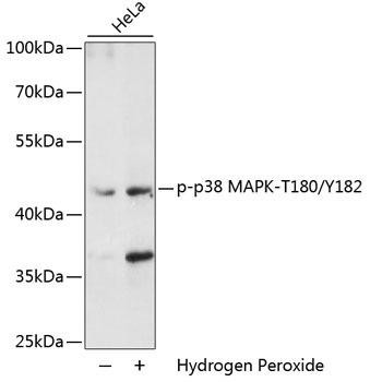
Phospho-p38 MAPK (Thr180/Tyr182) Antibody
Detects p38 phosphorylated on both activation-loop residues — the definitive marker of p38 activation.
View product →
Phospho-JNK (Thr183/Tyr185) ELISA Kit
Quantifies activated JNK without blotting, across the three common research species.
View product →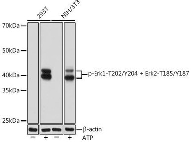
Phospho-ERK1/2 (Thr202/Tyr204, Thr185/Tyr187) Antibody
Reads the ERK branch at both isoforms’ dual phosphorylation sites.
View product →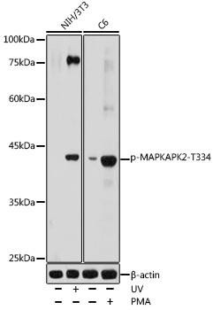
Phospho-MAPKAPK2 (Thr334) Antibody
MK2 is the p38 substrate that governs TNF mRNA stability — the step where p38 actually controls cytokine output.
View product →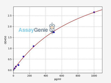
Human TNF-alpha ELISA Kit
Quantifies secreted TNF-alpha, the principal functional endpoint of p38-driven inflammation.
View product →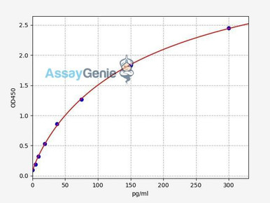
Human IL-6 ELISA Kit
Measures IL-6 release, the second major cytokine output of MAPK-driven inflammatory signalling.
View product →The three-tier cascade
The defining feature of MAPK signalling is its three-tier structure. Every branch follows the same pattern: a MAP kinase kinase kinase (MAP3K) phosphorylates a MAP kinase kinase (MAP2K), which phosphorylates the MAP kinase (MAPK), which then phosphorylates transcription factors and other substrates.
Three tiers rather than one is not redundancy. It amplifies, because each active kinase modifies many molecules of the next; it provides insulation, because scaffold proteins hold the tiers together so parallel cascades using shared components do not cross-talk indiscriminately; and it creates several independent points for regulation, feedback and drug intervention. It also means a stimulus can be sustained or transient depending on which tier feedback acts on — the same pathway components producing quite different response durations.
The three MAPK branches
Mammalian cells run three well-characterised branches. They share architecture but differ in their components, their preferred stimuli and their outputs.
| Branch | MAP3K | MAP2K | Typical stimuli and role |
|---|---|---|---|
| ERK1/2 | RAF family (A-RAF, B-RAF, C-RAF) | MEK1 and MEK2 | Growth factors and mitogens; proliferation, differentiation and survival. The branch targeted successfully in oncology. |
| JNK1, JNK2, JNK3 | MEKK1, ASK1, TAK1, MLK3 | MKK4 and MKK7 | Cytokines, oxidative and genotoxic stress; apoptosis, and AP-1-driven inflammatory transcription. |
| p38 (α, β, γ, δ) | TAK1, ASK1, MLK3, MEKK3/4 | MKK3 and MKK6 | Inflammatory cytokines, LPS, osmotic and oxidative stress; the principal inflammatory branch. |
The JNK and p38 branches are collectively described as stress-activated protein kinases, and they overlap substantially — TAK1 and ASK1 feed both. That overlap is one reason inhibiting a single branch often produces less effect than expected: the cell retains a parallel route to a similar output.
How MAPKs are activated
All MAPKs are switched on the same way: dual phosphorylation of a threonine and a tyrosine separated by one residue, the Thr-X-Tyr motif, within the activation loop. Both sites must be modified for full activity, and the responsible MAP2Ks are dual-specificity kinases capable of phosphorylating both.
- ERK1 and ERK2 are phosphorylated at Thr202/Tyr204 and Thr185/Tyr187 respectively.
- JNK is phosphorylated at Thr183/Tyr185.
- p38 is phosphorylated at Thr180/Tyr182.
This matters directly for experimental design. Total kinase levels barely change on stimulation, so measuring total p38 or total ERK tells you almost nothing about pathway activity. What changes is phosphorylation state at those specific residues, which is why phospho-specific antibodies raised against the exact dual sites are the standard readout, ideally alongside a total-protein blot to confirm the change is in phosphorylation rather than expression.
Routes from inflammatory receptors
The article’s central question is how inflammatory signals reach these cascades. Three routes dominate.
Toll-like receptors and IL-1 receptor
Recognition of microbial products by TLRs, or of IL-1 by IL-1R, recruits the adaptor MyD88, then IRAK kinases, then the ubiquitin ligase TRAF6. TRAF6 activates TAK1, which sits at the MAP3K tier and drives MKK3/6 to p38 and MKK4/7 to JNK. TAK1 also activates the IKK complex and hence NF-kappa-B, which is why these two pathways are engaged together and why their outputs are difficult to separate experimentally.
TNF receptor 1
TNF-alpha binding TNFR1 assembles a complex recruiting TRAF2 and RIP1, engaging MEKK1 and ASK1 at the MAP3K tier and thence JNK and p38. This creates a feed-forward loop: TNF activates MAPK signalling, and MAPK signalling promotes further TNF production, which is part of why inflammation becomes self-sustaining.
Oxidative stress
Reactive oxygen species oxidise thioredoxin, releasing ASK1 from inhibition. ASK1 then activates both JNK and p38, linking the redox state of the cell directly to inflammatory signalling — the route by which oxidative stress and inflammation reinforce one another.
Control of TNF-alpha and IL-6
MAPK signalling regulates cytokine production at two levels, and the second is frequently overlooked.
Transcriptionally, JNK and p38 phosphorylate transcription factors including c-Jun and ATF2, which dimerise to form AP-1. AP-1 sites occur in the promoters of many inflammatory genes, including TNF and IL6, and AP-1 acts in concert with NF-kappa-B to drive their expression.
Post-transcriptionally, p38 acts through MK2 — MAPK-activated protein kinase 2. Activated p38 phosphorylates MK2 at Thr334; MK2 in turn phosphorylates tristetraprolin, a protein that binds AU-rich elements in the 3′ untranslated region of TNF mRNA and targets it for degradation. Phosphorylated tristetraprolin can no longer destabilise the transcript, so the mRNA persists and more TNF protein is made.
This is the mechanistically important point about p38 and inflammation: much of its control over TNF operates through mRNA stability rather than transcription. It explains why p38 inhibition reduces TNF protein more than TNF transcription, and why MK2 has been pursued as a target in its own right — on the reasoning that inhibiting the specific effector might avoid the toxicity of inhibiting p38 wholesale.
Switching the signal off
The original framing that MAPK signalling both promotes and limits inflammation is correct, and the mechanism is worth naming. Termination is active, not passive decay.
The principal off-switches are the dual-specificity phosphatases, or MAP kinase phosphatases, which remove phosphate from both the threonine and the tyrosine of the activation loop. MKP-1, encoded by DUSP1, is itself induced by p38 signalling and preferentially dephosphorylates p38 and JNK — a negative feedback loop in which the pathway switches on its own inhibitor. Loss of MKP-1 markedly amplifies cytokine responses to LPS.
Two consequences follow. Steady-state phosphorylation reflects the balance between kinase and phosphatase, so a change in signal can arise from altered phosphatase activity with no change in kinase activity at all. And because the feedback takes time to build, signal duration is itself informative — which is why a single timepoint is rarely adequate and time courses are the norm in this field.
Therapeutic targeting
MAPK signalling looked like an ideal anti-inflammatory target, and the outcome is instructive about the difference between a good rationale and a usable drug.
The p38 story
p38 inhibitors reached clinical trials for rheumatoid arthritis and other inflammatory conditions on strong preclinical evidence, and they did engage the target and lower inflammatory markers. What they did not do was produce durable clinical benefit. Responses tended to attenuate over weeks, and hepatotoxicity and central nervous system effects appeared in several programmes. Development in inflammatory disease was largely abandoned as a result.
The likely explanations are informative in themselves: p38 also drives the MKP-1 feedback loop, so inhibiting it removes an off-switch as well as an on-switch; parallel routes through JNK and NF-kappa-B remain available; and p38 is required in many normal tissues, which narrows the therapeutic window.
Where the pathway has succeeded
The contrast with oncology is stark. Inhibitors of the ERK branch — BRAF inhibitors at the MAP3K tier, MEK inhibitors at the MAP2K tier — are approved and effective, particularly in combination for BRAF-mutant melanoma. The difference is that those tumours carry an activating mutation that makes them dependent on the pathway, whereas inflammatory cells retain alternatives. A pathway is druggable when the target cell has no way around it.
Current directions
Interest has shifted to targeting further down or further up: MK2, on the reasoning that hitting the specific effector of TNF mRNA stability may separate efficacy from p38-wide toxicity; and TAK1 or ASK1 at the MAP3K tier for greater stimulus selectivity. Meanwhile the approved anti-inflammatory drugs that work — TNF biologics and JAK inhibitors — act outside the MAPK cascade entirely, which is worth remembering when reading enthusiastic preclinical work.
Measuring pathway activity
Because activity is a phosphorylation state, experimental design follows a few consistent rules.
- Measure phospho, and normalise to total. Blot or assay the dual activation-loop sites, and run total kinase alongside so an apparent increase is not just higher expression.
- Take a time course. MAPK phosphorylation typically peaks within 15 to 60 minutes and resolves as phosphatases are induced. A single late timepoint can miss the response entirely.
- Read a downstream substrate as well. Phospho-MK2 confirms that active p38 is doing something, and guards against a phospho-p38 signal that reflects antibody cross-reactivity rather than function.
- Finish with a functional endpoint. Secreted TNF-alpha and IL-6 by ELISA are what the pathway output actually means; phosphorylation alone does not establish that cytokine production changed.
- Handle lysates properly. Include phosphatase inhibitors, keep samples cold and avoid freeze-thaw — phospho-epitopes are lost easily, and a negative result is often a handling artefact.
- Use pathway inhibitors as controls. Loss of the downstream signal in the presence of a specific inhibitor is far stronger evidence than a correlation between stimulus and phosphorylation.
References
- Kyriakis JM, Avruch J. Mammalian MAPK signal transduction pathways activated by stress and inflammation. Physiological Reviews, 2012;92(2):689–737.
- Arthur JSC, Ley SC. Mitogen-activated protein kinases in innate immunity. Nature Reviews Immunology, 2013;13(9):679–692.
- Wagner EF, Nebreda ÁR. Signal integration by JNK and p38 MAPK pathways in cancer development. Nature Reviews Cancer, 2009;9(8):537–549.
- Dhillon AS, Hagan S, Rath O, Kolch W. MAP kinase signalling pathways in cancer. Oncogene, 2007;26(22):3279–3290.
- Rincón M, Davis RJ. Regulation of the immune response by stress-activated protein kinases. Immunological Reviews, 2009;228(1):212–224.
- Zhang W, Liu HT. MAPK signal pathways in the regulation of cell proliferation in mammalian cells. Cell Research, 2002;12(1):9–18.
Choosing phospho antibodies
Phospho-specific antibodies against the activation-loop sites of p38, JNK and ERK, phospho-MK2 for downstream confirmation, and TNF-alpha and IL-6 ELISA kits for functional endpoints.
Browse phospho antibodies & kits →Frequently asked questions
What are the three MAPK pathways?
ERK1/2, JNK1-3 and p38. Each runs a three-tier cascade with its own MAP3Ks and MAP2Ks: RAF to MEK1/2 for ERK, several MAP3Ks to MKK4/7 for JNK, and TAK1 or ASK1 to MKK3/6 for p38. JNK and p38 are collectively the stress-activated kinases.
How do MAPKs get activated?
By dual phosphorylation of a threonine and tyrosine two residues apart in the activation loop — Thr180/Tyr182 for p38, Thr183/Tyr185 for JNK, Thr202/Tyr204 for ERK1. Both residues must be phosphorylated, which is why phospho-specific antibodies target the dual site.
Why measure phospho-MAPK rather than total MAPK?
Because total levels change very little on stimulation. Activity is entirely a matter of phosphorylation state, so a total blot will look flat while the pathway is strongly activated. Run both, and interpret phospho relative to total.
How does p38 control TNF-alpha production?
Substantially through mRNA stability rather than transcription. p38 phosphorylates MK2, which phosphorylates tristetraprolin, the protein that would otherwise degrade TNF mRNA via its AU-rich elements. Phosphorylated tristetraprolin cannot destabilise the transcript, so more TNF is produced.
How is MAPK signalling switched off?
By dual-specificity phosphatases such as MKP-1, which strip both phosphates from the activation loop. MKP-1 is itself induced by p38, forming a negative feedback loop — so the pathway switches on its own inhibitor, and signal duration depends on that balance.
Why did p38 inhibitors fail in rheumatoid arthritis?
They engaged the target and reduced inflammatory markers, but benefit was not durable and toxicity emerged. Contributing factors include loss of the MKP-1 feedback brake, parallel signalling through JNK and NF-kappa-B, and p38 being required in normal tissue.
Are any MAPK-pathway drugs approved?
Yes, but in oncology rather than inflammation — BRAF and MEK inhibitors targeting the ERK branch, especially combined in BRAF-mutant melanoma. Those tumours depend on the pathway, whereas inflammatory cells retain alternative routes.
Tehreem Ali completed her MS in Bioinformatics and conducted her research work at the IOMM lab at GCUF, Pakistan.
Recent Posts
-
Suspected Plague Death at a Siberian Anti-Plague Institute: What Researchers Should Know
Infectious Disease · News AnalysisSuspected Plague Death at a Siberian Anti-Plague Institute: What …6th Oct 2026 -
LPS ELISA Troubleshooting: The Importance of Sample Nature
Quick answer A researcher measuring circulating lipopolysaccharide (LPS) in human serum …25th Sep 2026 -

Sekundärantikörper wählen: Die Regel, die im Labor wirklich zählt
Sekundärantikörper wählen: Die Regel, die im Labor wirklich zählt Wählen Sie einen Sekundärantikörp …24th Sep 2026