The [KO Validated] CSDE1 Antibody (CAB5941) is a high-quality antibody developed for reliable detection and analysis of target proteins. This antibody, produced in rabbits, exhibits high affinity and specificity for CSDE1 in human samples, making it suitable for use in techniques such as Western blotting. By binding to the CSDE1 protein, this antibody facilitates the detection and analysis of CSDE1 in diverse cell types, making it ideal for investigations in molecular biology and cancer research.
This antibody is validated for use in WB, IF/ICC, IP, ELISA applications and has demonstrated reactivity against Human, Mouse samples.
Product Name:
[KO Validated] CSDE1 Antibody
SKU:
CAB5941
Size:
20μL, 100μL
Reactivity:
Human, Mouse
Conjugate:
Unconjugated
Immunogen:
Recombinant protein (or fragment).This information is considered to be commercially sensitive.
0.5μg-4μg antibody for 200μg-400μg extracts of whole cells
ELISA
Recommended starting concentration is 1 μg/mL. Please optimize the concentration based on your specific assay requirements.
Synonyms:
UNR, D1S155E, E1
Positive Sample:
HeLa
Cellular Localization:
Cytoplasm.
Calculated MW:
89kDa
Observed MW:
89kDa
Enables RNA stem-loop binding activity. Involved in IRES-dependent viral translational initiation; nuclear-transcribed mRNA catabolic process, no-go decay; and stress granule assembly. Located in Golgi apparatus; cytosol; and plasma membrane. Part of CRD-mediated mRNA stability complex.
Purification Method
Affinity purification
Gene ID
7812
RRID
AB_2716822
Buffer Information
Store at -20℃. Avoid freeze / thaw cycles. Buffer: PBS containing 50% glycerol, preserved with proclin300 or sodium azide, pH 7.3.
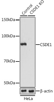
Western blot analysis of lysates from wild type (WT) and CSDE1 knockout (KO) HeLa cells, using [KO Validated] CSDE1 Rabbit pAb (CAB5941) at 1:1000 dilution. Secondary antibody: HRP-conjugated Goat anti-Rabbit IgG (H+L) (CABS014) at 1:10000 dilution. Lysates/proteins: 25μg per lane. Blocking buffer: 3% nonfat dry milk in TBST. Detection: ECL Basic Kit (AbGn00020). Exposure time: 15s.
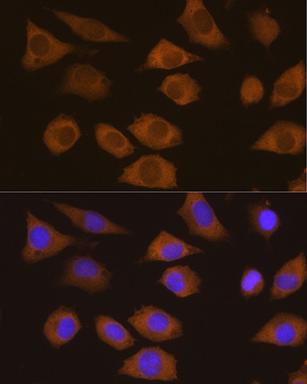
Immunofluorescence analysis of L929 cells using CSDE1 Rabbit pAb (CAB5941) at dilution of 1:100. Secondary antibody: Cy3-conjugated Goat anti-Rabbit IgG (H+L) (CABS007) at 1:500 dilution. Blue: DAPI for nuclear staining.