The EPHA2 Antibody (CAB7183) is a high-quality antibody developed for reliable detection and analysis of target proteins. This antibody, produced in rabbits, is highly specific to human samples and is suitable for use in Western blotting and immunohistochemistry applications.EphA2 plays a critical role in cancer progression and metastasis, making it a promising target for cancer research. By using the EphA2 Polyclonal Antibody, researchers can detect and analyze EphA2 expression in different cell types, providing insights into its function in cancer biology.
This antibody is validated for use in WB, ELISA applications and has demonstrated reactivity against Human, Mouse, Rat samples.
Product Name:
EPHA2 Antibody
SKU:
CAB7183
Size:
20μL, 100μL
Reactivity:
Human, Mouse, Rat
Conjugate:
Unconjugated
Immunogen:
Recombinant protein (or fragment).This information is considered to be commercially sensitive.
Recommended starting concentration is 1 μg/mL. Please optimize the concentration based on your specific assay requirements.
Synonyms:
ECK, CTPA, ARCC2, CTPP1, CTRCT6, EPHA2
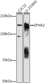
Positive Sample:
C2C12, U-251MG
Cellular Localization:
Cell Junction, Cell Membrane, Cell Projection, Single-Pass Type I Membrane Protein, Focal Adhesion, Lamellipodium Membrane, Ruffle Membrane.
Calculated MW:
108kDa
Observed MW:
125kDa
This gene belongs to the ephrin receptor subfamily of the protein-tyrosine kinase family. EPH and EPH-related receptors have been implicated in mediating developmental events, particularly in the nervous system. Receptors in the EPH subfamily typically have a single kinase domain and an extracellular region containing a Cys-rich domain and 2 fibronectin type III repeats. The ephrin receptors are divided into 2 groups based on the similarity of their extracellular domain sequences and their affinities for binding ephrin-A and ephrin-B ligands. This gene encodes a protein that binds ephrin-A ligands. Mutations in this gene are the cause of certain genetically-related cataract disorders.
Purification Method
Affinity purification
Gene ID
1969
RRID
AB_2767733
Buffer Information
Store at -20℃. Avoid freeze / thaw cycles. Buffer: PBS containing 50% glycerol, preserved with proclin300 or sodium azide, pH 7.3.
Western blot analysis of various lysates using EPHA2 Rabbit pAb (CAB7183) at 1:1000 dilution. Secondary antibody: HRP-conjugated Goat anti-Rabbit IgG (H+L) (CABS014) at 1:10000 dilution. Lysates/proteins: 25μg per lane. Blocking buffer: 3% nonfat dry milk in TBST. Detection: ECL Enhanced Kit (AbGn00021). Exposure time: 180s.