The [KO Validated] ERK2 Monoclonal Antibody (CAB19630) is a high-quality antibody developed for reliable detection and analysis of target proteins. This antibody, raised in rabbits, is rigorously validated for knockout (KO) specificity and is highly reactive with samples from various species, including human, mouse, and rat.ERK2, also known as extracellular signal-regulated kinase 2, plays a crucial role in cellular processes such as cell proliferation, differentiation, and survival. Dysregulation of the ERK2 pathway has been linked to various diseases, including cancer, neurodegenerative disorders, and inflammatory conditions.
This antibody is validated for use in WB, IHC-P, ELISA applications and has demonstrated reactivity against Human, Mouse, Rat samples.
Product Name:
[KO Validated] ERK2 Monoclonal Antibody
SKU:
CAB19630
Size:
20μL, 100μL
Reactivity:
Human, Mouse, Rat
Clone Number:
ARC51159
Conjugate:
Unconjugated
Immunogen:
Synthetic peptide. This information is considered to be commercially sensitive.
Cytoplasm, Nucleus, Centrosome, Cytoskeleton, Microtubule Organizing Center, Spindle.
Calculated MW:
41kDa
Observed MW:
41kDa
This gene encodes a member of the MAP kinase family. MAP kinases, also known as extracellular signal-regulated kinases (ERKs), act as an integration point for multiple biochemical signals, and are involved in a wide variety of cellular processes such as proliferation, differentiation, transcription regulation and development. The activation of this kinase requires its phosphorylation by upstream kinases. Upon activation, this kinase translocates to the nucleus of the stimulated cells, where it phosphorylates nuclear targets. One study also suggests that this protein acts as a transcriptional repressor independent of its kinase activity. The encoded protein has been identified as a moonlighting protein based on its ability to perform mechanistically distinct functions. Two alternatively spliced transcript variants encoding the same protein, but differing in the UTRs, have been reported for this gene.
Purification Method
Affinity purification
Gene ID
5594
RRID
AB_2862707
Buffer Information
Store at -20℃. Avoid freeze / thaw cycles. Buffer: PBS containing 50% glycerol and 0.05% BSA, preserved with proclin300 or sodium azide, pH 7.3.
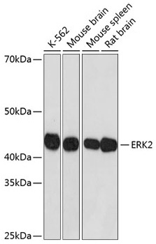
Western blot analysis of extracts of various cell lines using ERK2 Rabbit mAb (CAB19630) at 1:1000 dilution incubated overnight at 4℃. Secondary antibody: HRP Goat Anti-Rabbit IgG (H+L) (CABS014) at 1:10000 dilution. Lysates/proteins: 25 μg per lane. Blocking buffer: 3% nonfat dry milk in TBST. Detection: ECL Basic Kit (AbGn00020). Exposure time: 1 s.
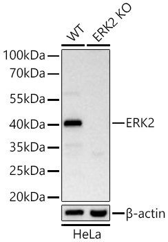
Western blot analysis of lysates from wild type (WT) and ERK2 knockout (KO) HeLa cells using ERK2 Rabbit mAb (CAB19630) at 1:2000 dilution incubated at room temperature for 1.5 hours. Secondary antibody: HRP-conjugated Goat anti-Rabbit IgG (H+L) (CABS014) at 1:10000 dilution. Lysates/proteins: 25 μg per lane. Blocking buffer: 3% nonfat dry milk in TBST. Detection: ECL Basic Kit (AbGn00020). Exposure time: 45 s.
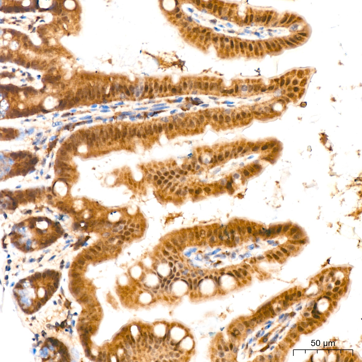
Immunohistochemistry analysis of paraffin-embedded Mouse intestin tissue using ERK2 Rabbit mAb (CAB19630) at a dilution of 1:200 (40x lens). High pressure antigen retrieval performed with 0.01M Citrate Buffer (pH 6.0) prior to IHC staining.
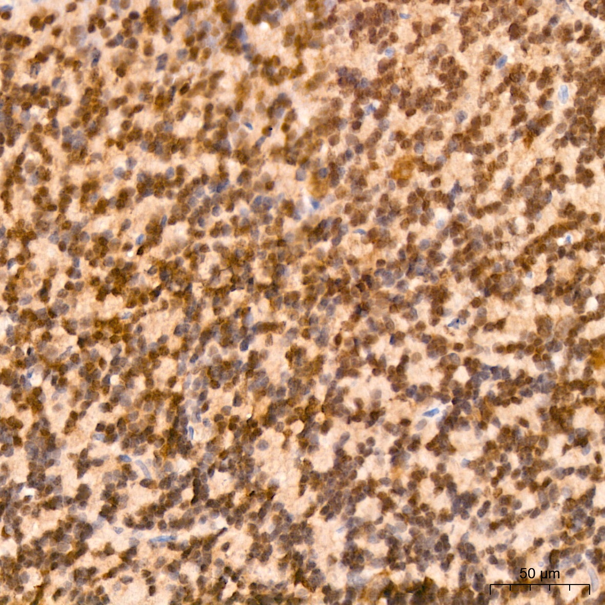
Immunohistochemistry analysis of paraffin-embedded Rat brain tissue using ERK2 Rabbit mAb (CAB19630) at a dilution of 1:200 (40x lens). High pressure antigen retrieval performed with 0.01M Citrate Buffer (pH 6.0) prior to IHC staining.