The [KO Validated] TMPO Antibody (CAB2534) is a high-quality antibody developed for reliable detection and analysis of target proteins. This antibody, produced in rabbits, is specifically designed for use in Western blot applications and is highly reactive with human samples.TMPO, also known as lamina-associated polypeptide 2, plays a crucial role in maintaining nuclear architecture and regulating gene expression. Aberrant expression or mutations in TMPO have been linked to diseases such as cancer and muscular dystrophy, making it an important target for further investigation.
This antibody is validated for use in WB, IHC-P, IF/ICC, ELISA applications and has demonstrated reactivity against Human, Mouse, Rat samples.
Product Name:
[KO Validated] TMPO Antibody
SKU:
CAB2534
Size:
20μL, 100μL
Reactivity:
Human, Mouse, Rat
Conjugate:
Unconjugated
Immunogen:
Recombinant protein (or fragment).This information is considered to be commercially sensitive.
Recommended starting concentration is 1 μg/mL. Please optimize the concentration based on your specific assay requirements.
Synonyms:
TP, LAP2, CMD1T, LEMD4, PRO0868, TMPO
Positive Sample:
HeLa
Cellular Localization:
Chromosome, Nucleus.
Calculated MW:
38kDa/50kDa/75kDa
Observed MW:
80kDa/51kDa
Through alternative splicing, this gene encodes several distinct LEM domain containing protein isoforms. LEM domain proteins include inner nuclear membrane and intranuclear proteins, and are involved in a variety of cellular functions including gene expression, chromatin organization, and replication and cell cycle control. The encoded alpha isoform is broadly diffuse in the nucleus and contains a lamin binding domain, while the beta and gamma isoforms are localized to the nuclear membrane and contain an HDAC3 interaction domain. The distinct isoforms may compete with each other when acting to chaperone other proteins and regulate transcription.
Purification Method
Affinity purification
Gene ID
7112
RRID
AB_2863011
Buffer Information
Store at -20℃. Avoid freeze / thaw cycles. Buffer: PBS containing 50% glycerol, preserved with proclin300 or sodium azide, pH 7.3.
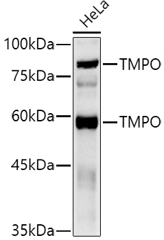
Western blot analysis of lysates from HeLa cells, using TMPO Rabbit pAb (CAB2534) at 1:500 dilution. Secondary antibody: HRP-conjugated Goat anti-Rabbit IgG (H+L) (CABS014) at 1:10000 dilution. Lysates/proteins: 25μg per lane. Blocking buffer: 3% nonfat dry milk in TBST. Detection: ECL Basic Kit (AbGn00020). Exposure time: 30s.
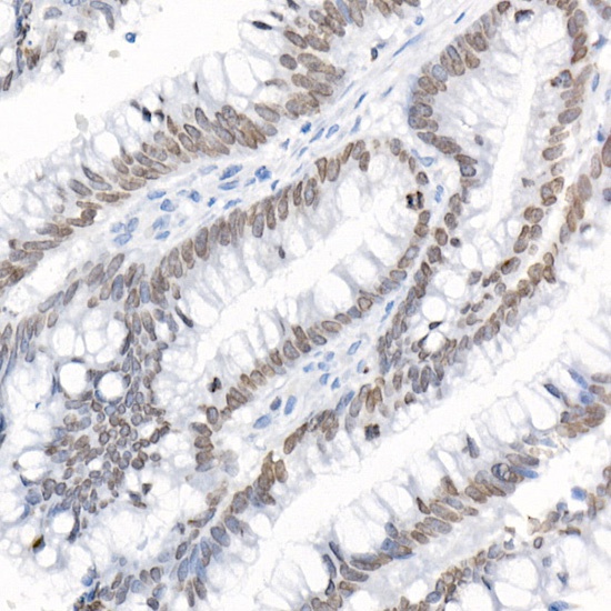
Immunohistochemistry analysis of paraffin-embedded Human colon carcinoma using TMPO Rabbit pAb (CAB2534) at dilution of 1:20 (40x lens). High pressure antigen retrieval performed with 0.01M Citrate buffer (pH 6.0) prior to IHC staining.
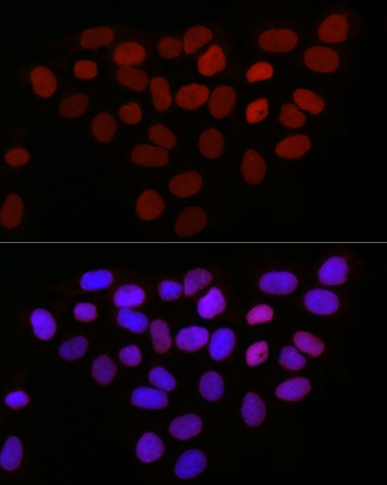
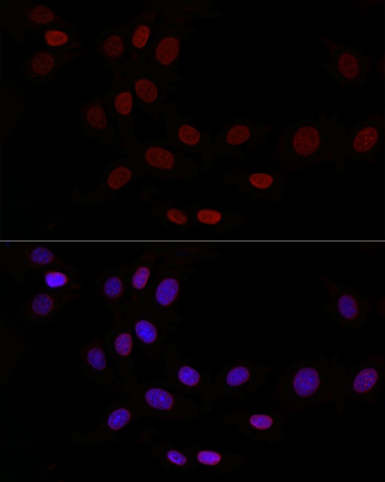
Immunofluorescence analysis of HeLa cells using TMPO Rabbit pAb (CAB2534) at dilution of 1:50 (40x lens). Secondary antibody: Cy3-conjugated Goat anti-Rabbit IgG (H+L) (CABS007) at 1:500 dilution. Blue: DAPI for nuclear staining.
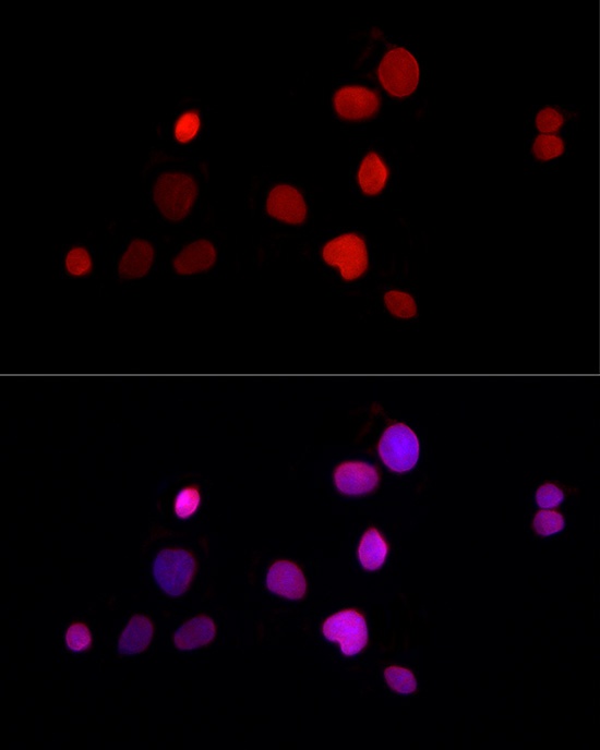
Immunofluorescence analysis of HepG2 cells using TMPO Rabbit pAb (CAB2534) at dilution of 1:50 (40x lens). Secondary antibody: Cy3-conjugated Goat anti-Rabbit IgG (H+L) (CABS007) at 1:500 dilution. Blue: DAPI for nuclear staining.
Immunofluorescence analysis of NIH/3T3 cells using TMPO Rabbit pAb (CAB2534) at dilution of 1:50 (40x lens). Secondary antibody: Cy3-conjugated Goat anti-Rabbit IgG (H+L) (CABS007) at 1:500 dilution. Blue: DAPI for nuclear staining.