The Phospho-ABL1-Y412 Antibody (CABP1060) is a high-quality antibody developed for reliable detection and analysis of target proteins. This antibody, raised in rabbits, specifically detects the phosphorylated form of ABL1 at tyrosine 412 and has been validated for use in Western blot applications. It enables the detection and analysis of phosphorylated ABL1 in human samples, providing insights into the regulation of cell signaling pathways.ABL1 is a key player in various cellular processes, including cell proliferation, differentiation, and migration.
This antibody is validated for use in WB, IHC-P, ELISA applications and has demonstrated reactivity against Human, Mouse, Rat samples.
Product Name:
Phospho-ABL1-Y412 Antibody
SKU:
CABP1060
Size:
20μL, 100μL
Reactivity:
Human, Mouse, Rat
Immunogen:
Synthetic peptide. This information is considered to be commercially sensitive.
Sequence:
DTYT A
Tested Applications:
WBIHC-PELISA
Recommended Dilution:
WB
1:500 - 1:2000
IHC-P
1:50 - 1:200
ELISA
Recommended starting concentration is 1 μg/mL. Please optimize the concentration based on your specific assay requirements.
This gene is a protooncogene that encodes a protein tyrosine kinase involved in a variety of cellular processes, including cell division, adhesion, differentiation, and response to stress. The activity of the protein is negatively regulated by its SH3 domain, whereby deletion of the region encoding this domain results in an oncogene. The ubiquitously expressed protein has DNA-binding activity that is regulated by CDC2-mediated phosphorylation, suggesting a cell cycle function. This gene has been found fused to a variety of translocation partner genes in various leukemias, most notably the t(9;22) translocation that results in a fusion with the 5' end of the breakpoint cluster region gene (BCR; MIM:151410). Alternative splicing of this gene results in two transcript variants, which contain alternative first exons that are spliced to the remaining common exons.
Purification Method
Affinity purification
Gene ID
25
RRID
AB_2863932
Buffer Information
Store at -20℃. Avoid freeze / thaw cycles. Buffer: PBS with 0.01% thimerosal,50% glycerol,pH7.3.
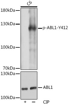
Western blot analysis of lysates from C6 cells, using Phospho-c-Abl-Y412 Rabbit pAb (CAB0282). C6 cells were treated by CIP(20uL/400ul) at 37℃ for 1 hour. Secondary antibody: HRP-conjugated Goat anti-Rabbit IgG (H+L) (CABS014) at 1:10000 dilution. Lysates/proteins: 25μg per lane. Blocking buffer: 3% BSA. Detection: ECL Basic Kit (AbGn00020). Exposure time: 3s.
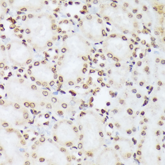
Immunohistochemistry analysis of paraffin-embedded Rat kidney using Phospho-c-Abl-Y412 Rabbit pAb (CABP1060) at dilution of 1:100 (40x lens). Microwave antigen retrieval performed with 0.01M Tris/EDTA Buffer (pH 9.0) prior to IHC staining.
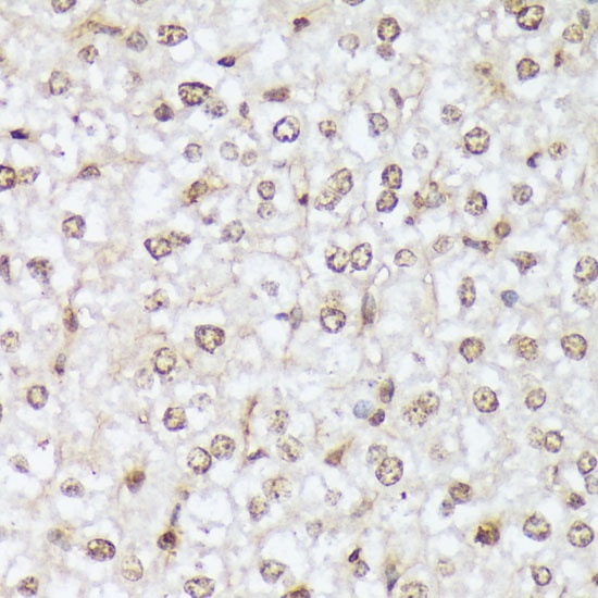
Immunohistochemistry analysis of paraffin-embedded Mouse liver using Phospho-c-Abl-Y412 Rabbit pAb (CABP1060) at dilution of 1:100 (40x lens). Microwave antigen retrieval performed with 0.01M Tris/EDTA Buffer (pH 9.0) prior to IHC staining.