XRCC6/XRCC5 Antibody is a premium polyclonal that offers outstanding performance and reliability for demanding research applications. Rigorously validated for ELISA, WB, IHC, IF, this antibody ensures consistent, reproducible results across multiple experimental platforms. Demonstrates excellent reactivity with Human, Mouse, Rat samples, providing researchers with confidence in cross-species compatibility. Conveniently packaged in 100ul format to meet your experimental needs. For optimal performance, store at -20°C or -80°C and maintains stability for 12 months. Backed by rigorous quality control testing to ensure superior performance in your critical research applications.
Product Name:
XRCC6/XRCC5 Antibody
SKU:
PACO23829
Size:
100μl
Host Species:
Rabbit
Reactivity:
Human, Mouse, Rat
Immunogen:
Synthesized peptide derived from Human Ku70/80.
Immunogen Species:
Homo sapiens (Human)
Uniprot No:
P12956
Form:
Rabbit IgG in phosphate buffered saline (without Mg2+ and Ca2+), pH 7.4, 150mM NaCl, 0.02% sodium azide and 50% glycerol.
Tested Applications:
ELISAWBIHCIF
Recommended Dilution:
Application
Recommended Dilution
WB
1:500-1:3000
IHC
1:50-1:100
IF
1:100-1:500
Synonyms:
5''-deoxyribose-5-phosphate lyase Ku70 antibody, 5''-dRP lyase Ku70 antibody, 70 kDa subunit of Ku antigen antibody, ATP dependent DNA helicase 2 subunit 1 antibody, ATP dependent DNA helicase II 70 kDa subunit antibody, ATP-dependent DNA helicase 2 subunit 1 antibody, ATP-dependent DNA helicase II 70 kDa subunit antibody, CTC box binding factor 75 kDa subunit antibody, CTC box-binding factor 75 kDa subunit antibody, CTC75 antibody, CTCBF antibody, DNA repair protein XRCC6 antibody, G22P1 antibody, Ku 70 antibody, Ku autoantigen p70 subunit antibody, Ku autoantigen, 70kDa antibody, Ku p70 anti...Read more
Target Names:
XRCC6/XRCC5
Purification:
The antibody was affinity-purified from rabbit antiserum by affinity-chromatography using epitope-specific immunogen.
Clonality:
Polyclonal
Conjugate:
Non-conjugated
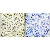
Immunohistochemical analysis of paraffin-embedded human breast carcinoma tissue using Ku70/80 antibody.
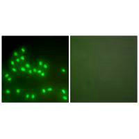
Immunofluorescence analysis of A549 cells, using Ku70/80 antibody.
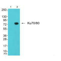
Western blot analysis of extracts from LOVO cells, using Ku70/80 antibody.
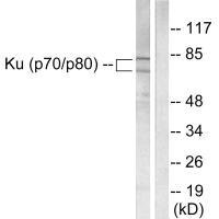
Western blot analysis of extracts from JK cells (Lane 2), using Ku80 antiobdy. The lane on the left is treated with synthesized peptide.


")


