The [KO Validated] ENO1 Antibody (CAB1033) is a high-quality antibody developed for reliable detection and analysis of target proteins. This antibody, raised in rabbits, is highly reactive with human samples and is validated for use in applications such as Western blotting and immunohistochemistry.Alpha-enolase is a multifunctional enzyme that not only catalyzes the conversion of 2-phosphoglycerate to phosphoenolpyruvate in glycolysis but also functions as a plasminogen receptor on the cell surface, leading to roles in cell migration and invasion.
This antibody is validated for use in WB, IHC-P, IF/ICC, IP, ELISA applications and has demonstrated reactivity against Human, Mouse samples.
Product Name:
[KO Validated] ENO1 Antibody
SKU:
CAB1033
Size:
20μL, 100μL
Reactivity:
Human, Mouse
Conjugate:
Unconjugated
Immunogen:
Recombinant protein (or fragment).This information is considered to be commercially sensitive.
0.5μg-4μg antibody for 200μg-400μg extracts of whole cells
ELISA
Recommended starting concentration is 1 μg/mL. Please optimize the concentration based on your specific assay requirements.
Synonyms:
NNE, PPH, MPB1, ENO1L1, ENO1-IT1, HEL-S-17, ENO1
Positive Sample:
HeLa, SH-SY5Y, MCF-7, Jurkat, NIH/3T3
Cellular Localization:
Cell Membrane, Cytoplasm, M Line, Nucleus, Myofibril, Sarcomere.
Calculated MW:
47kDa
Observed MW:
47kDa
This gene encodes alpha-enolase, one of three enolase isoenzymes found in mammals. Each isoenzyme is a homodimer composed of 2 alpha, 2 gamma, or 2 beta subunits, and functions as a glycolytic enzyme. Alpha-enolase in addition, functions as a structural lens protein (tau-crystallin) in the monomeric form. Alternative splicing of this gene results in a shorter isoform that has been shown to bind to the c-myc promoter and function as a tumor suppressor. Several pseudogenes have been identified, including one on the long arm of chromosome 1. Alpha-enolase has also been identified as an autoantigen in Hashimoto encephalopathy.
Purification Method
Affinity purification
Gene ID
2023
RRID
AB_2757874
Buffer Information
Store at -20℃. Avoid freeze / thaw cycles. Buffer: PBS with 0.09% sodium azide,50% glycerol,pH7.3.
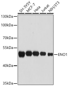
Western blot analysis of various lysates using ENO1 Rabbit pAb (CAB1033) at 1:3000 dilution incubated overnight at 4℃. Secondary antibody: HRP-conjugated Goat anti-Rabbit IgG (H+L) (CABS014) at 1:10000 dilution. Lysates/proteins: 25 μg per lane. Blocking buffer: 3% nonfat dry milk in TBST. Detection: ECL Basic Kit (AbGn00020) Exposure time: 1 s.
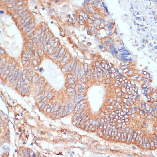
Immunohistochemistry analysis of paraffin-embedded Human colon carcinoma using ENO1 Rabbit pAb (CAB1033) at dilution of 1:100 (40x lens). Microwave antigen retrieval performed with 0.01M PBS Buffer (pH 7.2) prior to IHC staining.
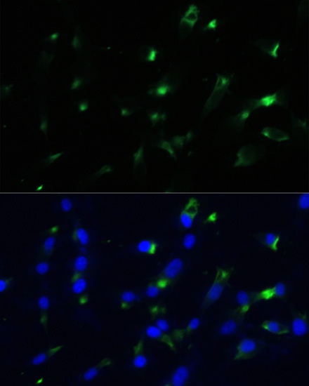
Immunofluorescence analysis of NIH-3T3 cells using ENO1 Rabbit pAb (CAB1033) at dilution of 1:100. Secondary antibody: Cy3-conjugated Goat anti-Rabbit IgG (H+L) (CABS007) at 1:500 dilution. Blue: DAPI for nuclear staining.
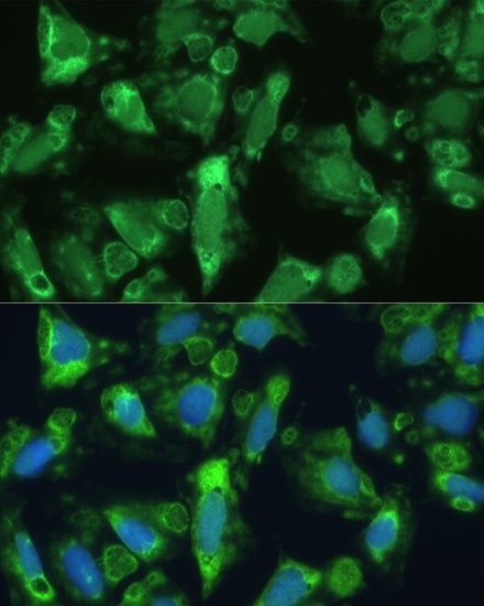
Immunofluorescence analysis of U-2 OS cells using ENO1 Rabbit pAb (CAB1033) at dilution of 1:100. Secondary antibody: Cy3-conjugated Goat anti-Rabbit IgG (H+L) (CABS007) at 1:500 dilution. Blue: DAPI for nuclear staining.
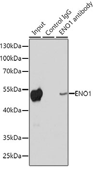
Immunoprecipitation analysis of 200 μg extracts of HeLa cells using 1 μg ENO1 Rabbit pAb (CAB1033). Western blot was performed from the immunoprecipitate using ENO1 antibody (CAB1033) at a dilution of 1:1000.