The SUPT5H/SPT5 Monoclonal Antibody (CAB19225) is a high-quality antibody developed for reliable detection and analysis of target proteins. This antibody, generated in rabbits, exhibits high specificity for human samples and has been validated for use in Western blot applications.Supt5h/Spt5 is a key component of the DSIF (DRB-sensitivity inducing factor) complex, which facilitates the elongation phase of RNA polymerase II transcription. Dysregulation of this process has been linked to various diseases, including cancer and developmental disorders.
This antibody is validated for use in WB, IHC-P, IF/ICC, IP, ELISA applications and has demonstrated reactivity against Human, Mouse, Rat samples.
Product Name:
SUPT5H/SPT5 Monoclonal Antibody
SKU:
CAB19225
Size:
20μL, 100μL
Reactivity:
Human, Mouse, Rat
Clone Number:
ARC2380
Conjugate:
Unconjugated
Immunogen:
Synthetic peptide. This information is considered to be commercially sensitive.
0.5μg-4μg antibody for 200μg-400μg extracts of whole cells
ELISA
Recommended starting concentration is 1 μg/mL. Please optimize the concentration based on your specific assay requirements.
Synonyms:
SPT5, SPT5H, Tat-CT1, SUPT5H/SPT5
Positive Sample:
HeLa,
Cellular Localization:
Nucleus.
Calculated MW:
121kDa
Observed MW:
150kDa
Enables enzyme binding activity and protein heterodimerization activity. Involved in positive regulation of macroautophagy; regulation of RNA metabolic process; and transcription elongation from RNA polymerase II promoter. Located in nucleoplasm. Part of DSIF complex.
Purification Method
Affinity purification
Gene ID
6829
Buffer Information
Store at -20℃. Avoid freeze / thaw cycles. Buffer: PBS containing 50% glycerol and 0.05% BSA, preserved with proclin300 or sodium azide, pH 7.3.
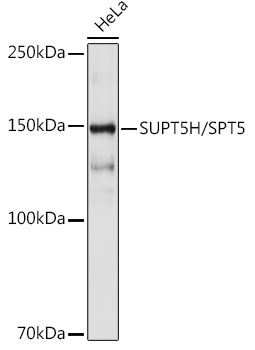
Western blot analysis of lysates from HeLa cells, using SUPT5H/SPT5 Rabbit mAb (CAB19225) at 1:1000 dilution. Secondary antibody: HRP-conjugated Goat anti-Rabbit IgG (H+L) (CABS014) at 1:10000 dilution. Lysates/proteins: 25μg per lane. Blocking buffer: 3% nonfat dry milk in TBST. Detection: ECL Basic Kit (AbGn00020). Exposure time: 10s.
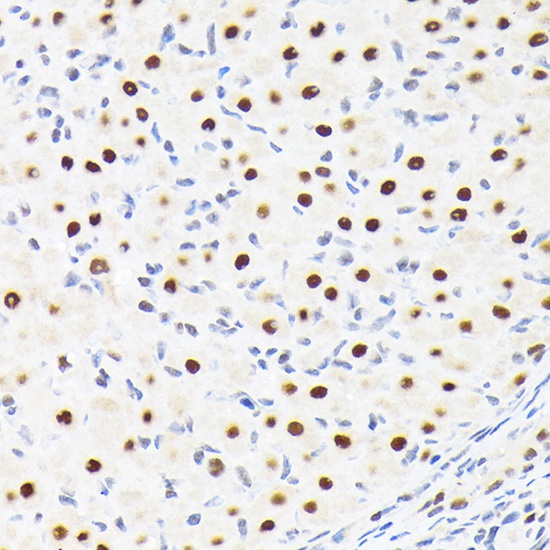
Immunohistochemistry analysis of paraffin-embedded Rat ovary using SUPT5H/SPT5 Rabbit mAb (CAB19225) at dilution of 1:100 (40x lens). Microwave antigen retrieval performed with 0.01M Tris/EDTA Buffer (pH 9.0) prior to IHC staining.
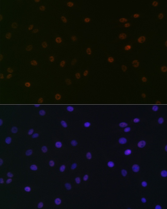
Immunofluorescence analysis of C6 cells using SUPT5H/SPT5 Rabbit mAb (CAB19225) at dilution of 1:100 (40x lens). Secondary antibody: Cy3-conjugated Goat anti-Rabbit IgG (H+L) (CABS007) at 1:500 dilution. Blue: DAPI for nuclear staining.
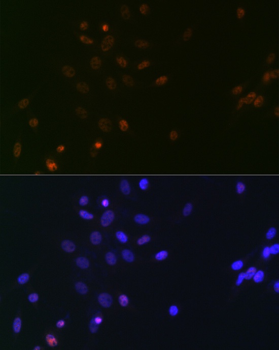
Immunofluorescence analysis of NIH/3T3 cells using SUPT5H/SPT5 Rabbit mAb (CAB19225) at dilution of 1:100 (40x lens). Secondary antibody: Cy3-conjugated Goat anti-Rabbit IgG (H+L) (CABS007) at 1:500 dilution. Blue: DAPI for nuclear staining.
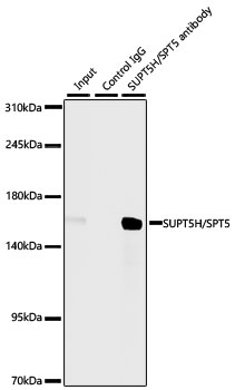
Immunoprecipitation of SUPT5H/SPT5 from 300 µg extracts of HeLa cells was performed using 3 µg of SUPT5H/SPT5 Rabbit mAb (CAB19225). Rabbit Control IgG (AC005) was used to precipitate the Control IgG sample. IP samples were eluted with 1X Laemmli Buffer. The Input lane represents 10% of the total input. Western blot analysis of immunoprecipitates was conducted using SUPT5H/SPT5 Rabbit mAb (CAB19225) at a dilution of 1:1000.


![Anti-SPT5 [R01-5C2] Monoclonal Antibody (AGMB02550)](https://cdn11.bigcommerce.com/s-h68l9z2lnx/images/stencil/590x590/products/273839/678057/anti-spt5-r01-5c2-monoclonal-antibody-agmb02550__22423.1773034578.jpg?c=2 "Anti-SPT5 [R01-5C2] Monoclonal Antibody (AGMB02550)")
![Anti-SPT5 [R03-2Q1] Monoclonal Antibody (AGMB03290)](https://cdn11.bigcommerce.com/s-h68l9z2lnx/images/stencil/590x590/products/274579/679907/anti-spt5-r03-2q1-monoclonal-antibody-agmb03290__56210.1773040360.jpg?c=2 "Anti-SPT5 [R03-2Q1] Monoclonal Antibody (AGMB03290)")


