
C-Reactive Protein (CRP): Function, Levels & What They Mean
C-Reactive Protein (CRP): Structure, Function and Clinical Use
CRP is the most widely measured marker of inflammation in medicine — fast to rise, fast to fall, and entirely non-specific about cause. This guide covers its pentameric structure and IL-6-driven synthesis, what it actually does in innate immunity, the reference ranges and the important distinction between standard and high-sensitivity assays, and how it is interpreted alongside procalcitonin and ESR.
Browse CRP assays →Key takeaways
- CRP is an acute-phase protein made by hepatocytes in response to IL-6, rising within hours of an inflammatory stimulus.
- It is a pentamer of five identical 206-amino-acid subunits arranged around a central pore, encoded by CRP on chromosome 1q23.2.
- Functionally it binds phosphocholine on damaged membranes and microbial surfaces, acting as an opsonin and activating complement through C1q.
- Its short half-life of 18 to 24 hours is what makes serial measurement useful — levels track the inflammatory process closely in both directions.
- CRP is sensitive but wholly non-specific: it indicates that inflammation is present, not where it is or what is causing it.
- Standard and high-sensitivity assays answer different questions — hs-CRP for cardiovascular risk stratification at low concentrations, standard CRP for infection and active inflammatory disease.
- Interpretation improves markedly when CRP is read alongside procalcitonin, which discriminates bacterial from viral infection far better than CRP alone.
CRP and acute-phase assay kits
CRP is rarely informative in isolation. The panel below pairs it with the cytokine that drives it, the marker that discriminates bacterial infection, and the second major acute-phase protein.
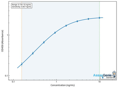
Human C-Reactive Protein (CRP) ELISA Kit
Quantifies CRP in serum and plasma — the core measurement for inflammatory status.
View product →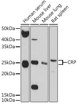
C-Reactive Protein Rabbit Polyclonal Antibody
Localises CRP in tissue, for work on local rather than circulating expression.
View product →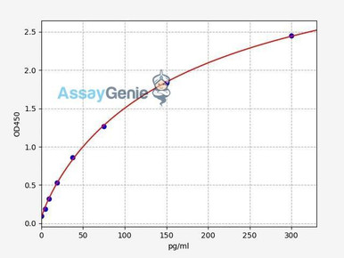
Human IL-6 ELISA Kit
The cytokine driving hepatic CRP synthesis; measuring both separates stimulus from response.
View product →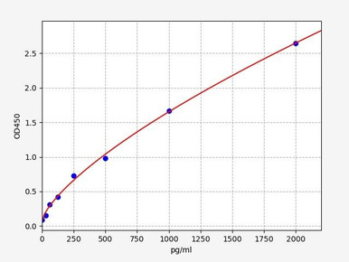
Human Procalcitonin ELISA Kit
Rises selectively in bacterial infection, so it addresses the question CRP cannot answer.
View product →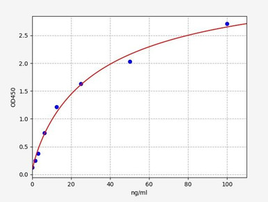
Human Serum Amyloid A ELISA Kit
The other major acute-phase protein, with a faster and larger dynamic response than CRP.
View product →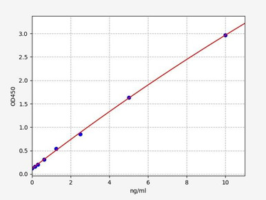
Rat CRP ELISA Kit (High Sensitivity)
High-sensitivity detection for preclinical inflammation models.
View product →What is C-reactive protein?
C-reactive protein is an acute-phase reactant — a plasma protein whose concentration rises sharply during inflammation. It is one of the earliest and largest responders, capable of increasing several hundred-fold from baseline, which is why it became the standard laboratory index of inflammatory activity.
The name is historical rather than descriptive. It was identified in the serum of patients with acute pneumococcal infection, where it reacted with the pneumococcal capsular C-polysaccharide. That reaction is now understood as binding to phosphocholine, a group present both in bacterial polysaccharides and on the surface of damaged host cells.
Gene and structure
CRP is encoded by the CRP gene at chromosome 1q23.2. Transcription is driven principally by IL-6, with IL-1 beta acting synergistically, which is why CRP concentration is effectively a downstream readout of cytokine activity rather than an independent variable.
The protein is a pentamer: five identical subunits of 206 amino acids, each around 23 kDa, held together non-covalently in a disc with a central pore. The two faces of the disc are functionally distinct — one binds phosphocholine in a calcium-dependent manner, the other engages C1q and Fc receptors. That arrangement is what allows a single molecule to recognise a target and recruit effectors simultaneously.
A detail worth knowing for research work: pentameric CRP can dissociate into monomeric CRP at inflamed tissue surfaces, and the monomeric form has distinct, largely pro-inflammatory activity. Standard clinical assays measure the pentameric form, so the two are not interchangeable and assay choice matters if the monomer is the target of interest.
How CRP is produced
The pathway is short and worth stating in order, because each step explains a feature of the marker’s behaviour.
An inflammatory stimulus — infection, trauma, surgery, tissue necrosis, malignancy or autoimmune activity — causes macrophages and adipocytes to release IL-6. IL-6 travels to the liver and binds receptors on hepatocytes, activating transcription of CRP. The protein is synthesised, secreted, and appears in plasma within four to six hours, peaking at around 48 hours. When the stimulus resolves, synthesis stops and concentration falls according to the 18 to 24 hour half-life.
Two consequences follow. Because clearance is constant and rapid, plasma CRP reflects current synthesis rate almost directly — there is no accumulation from previous days. And because adipose tissue produces IL-6, obesity raises baseline CRP independently of any acute illness, which is a recurring confounder in cardiovascular risk work.
Function in immunity
CRP is not merely a marker; it is an effector of innate immunity, functioning as a soluble pattern recognition molecule.
- Ligand recognition. CRP binds phosphocholine exposed on bacterial surfaces and on the membranes of apoptotic and damaged host cells, in a calcium-dependent manner.
- Opsonisation. Bound CRP is recognised by Fc gamma receptors on phagocytes, marking the target for phagocytosis by macrophages and neutrophils.
- Complement activation. CRP engages C1q and activates the classical pathway. Importantly, it activates the early components while recruiting regulators that limit progression to the terminal lytic complex — so it promotes clearance more than lysis.
- Clearance of dead cells. By binding apoptotic membranes and facilitating their uptake, CRP contributes to removing cellular debris without provoking autoimmunity.
- Modulation of inflammation. CRP influences cytokine production and leukocyte recruitment, with effects that are context-dependent and differ between the pentameric and monomeric forms.
Measuring CRP: standard and high-sensitivity
The two assay classes are frequently conflated, and choosing the wrong one wastes the measurement.
| Assay | Detection range | Question it answers |
|---|---|---|
| Standard CRP | Lower limit around 3–5 mg/L | Is there significant acute inflammation or infection, and is it resolving? |
| High-sensitivity CRP (hs-CRP) | Down to approximately 0.1 mg/L | Where does this person sit on the low-grade inflammation spectrum, for cardiovascular risk stratification? |
The distinction is one of analytical sensitivity, not of a different analyte — both measure the same pentameric protein. Standard assays cannot resolve differences in the 1 to 3 mg/L range where cardiovascular risk stratification operates, which is the entire reason hs-CRP exists. Conversely, using hs-CRP to monitor a patient with sepsis adds nothing, because concentrations are far above the range where the extra sensitivity matters.
In research settings, sandwich ELISA is the standard quantitative approach for serum and plasma. Check the stated dynamic range against the concentrations you expect, since a kit optimised for hs-CRP work will saturate in acute inflammatory samples and require dilution.
Reference ranges and interpretation
Units are a common source of confusion. CRP is usually reported in mg/L in most laboratories, but some report mg/dL, and the two differ tenfold — 3 mg/L equals 0.3 mg/dL. Always confirm which unit a result is in before comparing values.
| Concentration | Interpretation | Typical context |
|---|---|---|
| Below 1 mg/L | Low | hs-CRP: lower cardiovascular risk category |
| 1–3 mg/L | Intermediate | hs-CRP: average cardiovascular risk category |
| Above 3 mg/L | Raised low-grade inflammation | hs-CRP: higher cardiovascular risk category; also seen with obesity |
| 10–40 mg/L | Mild to moderate inflammation | Viral infection, mild inflammatory conditions, post-operative recovery |
| 40–200 mg/L | Marked inflammation | Bacterial infection, active autoimmune disease, significant tissue injury |
| Above 200 mg/L | Severe | Severe bacterial infection, sepsis, extensive trauma or burns |
These bands are indicative rather than diagnostic thresholds, and they overlap substantially. A meaningful proportion of apparently healthy people sit above 3 mg/L for reasons including adiposity, smoking and genetic variation — which is why the cardiovascular categories describe populations rather than individuals. Factors that shift CRP independently of acute illness include age, sex (women tend slightly higher), obesity, smoking, exercise, hormonal status, statins and NSAIDs, chronic kidney disease and rheumatoid arthritis.
The trend matters more than the single value. Because of the short half-life, serial measurements track a process far better than one reading, and a falling CRP is often the most useful evidence that treatment is working. Nothing here is clinical guidance; interpretation belongs with the treating clinician.
CRP versus ESR and procalcitonin
CRP is one of three commonly used inflammatory indices, and their differences are practical rather than academic.
| Marker | What it measures | Kinetics | Best used for |
|---|---|---|---|
| CRP | Hepatic acute-phase protein concentration | Rises in 4–6 h, peaks ~48 h, half-life 18–24 h | Detecting and tracking inflammation of any cause; monitoring response |
| ESR | Rate of erythrocyte sedimentation, reflecting fibrinogen and immunoglobulin | Rises and falls over days to weeks | Chronic conditions where a slower index is wanted, such as polymyalgia rheumatica |
| Procalcitonin | Precursor peptide induced by bacterial products | Rises in 3–6 h, half-life ~24 h | Discriminating bacterial from viral infection; antibiotic stewardship decisions |
The key contrast is specificity. CRP and ESR both rise in essentially any inflammatory state, so neither identifies the cause. Procalcitonin is comparatively selective for bacterial infection, remaining low in most viral illness, which is why it is used to support decisions about starting or stopping antibiotics where CRP alone is ambiguous. CRP and ESR frequently diverge because their kinetics differ — a resolving process shows falling CRP while ESR remains raised, and that discordance is informative rather than contradictory.
Clinical associations
Raised CRP accompanies a wide range of conditions. The table separates them by how the marker is typically used rather than listing them undifferentiated.
| Condition | CRP behaviour | How it is used |
|---|---|---|
| Bacterial infection | Marked rise, often 50–100 mg/L within 4–6 hours and higher in severe disease | Detecting infection and tracking response to antibiotics |
| Viral infection | Usually modest rise | Poor discriminator alone — procalcitonin is more informative |
| Rheumatoid arthritis | Raised with active disease | Assessing disease activity and treatment response; incorporated into composite activity scores |
| Systemic lupus erythematosus | Often only modestly raised even in active flare | Notable exception — a normal CRP does not exclude active SLE, though it rises with intercurrent infection |
| Inflammatory bowel disease | Raised in active Crohn’s disease; less reliably in ulcerative colitis | Monitoring activity, usually with faecal calprotectin |
| Cardiovascular disease | Low-grade elevation in the hs-CRP range | Risk stratification alongside lipids and conventional risk factors |
| Obesity and metabolic syndrome | Chronic mild elevation from adipose IL-6 | A confounder to account for rather than a finding to act on |
| Obstructive sleep apnoea | Mild chronic elevation | Reflects low-grade inflammation contributing to cardiovascular risk |
The SLE row is the clinically important exception and is often missed: unlike most inflammatory diseases, active lupus frequently produces only a modest CRP rise, so a normal value cannot be used to rule out a flare. A markedly raised CRP in a lupus patient more often indicates intercurrent infection than disease activity.
Genetic variation also contributes. Carriers of the APOE4 allele have been reported to show higher CRP concentrations, one of several associations linking the variant to inflammatory and cardiovascular risk.
CRP and cancer
Elevated CRP has been observed across many malignancies including lung, colorectal, breast, pancreatic and ovarian cancer, and the relationship runs in both directions.
As a prognostic indicator. A raised CRP at diagnosis is associated with more advanced stage and poorer outcome in several cancers, and it forms part of composite prognostic indices in some settings. It is not a diagnostic test — it cannot detect or localise a tumour — but it carries prognostic information once a diagnosis exists, and a fall during treatment may reflect response.
As a reflection of tumour-associated inflammation. The tumour microenvironment releases cytokines including IL-6, which drives hepatic CRP synthesis. Since chronic inflammation supports proliferation, angiogenesis, invasion and immune evasion, raised CRP is a visible correlate of a process that contributes to progression rather than a bystander.
Prospective cohort data on colorectal cancer illustrate both the signal and its limits: individuals who later developed colorectal cancer had higher mean CRP than those who did not, yet values in both groups largely sat within the conventional normal range. The difference is real at population level and useless at individual level — which is the general shape of CRP as a cancer-risk marker, and consistent with the separate observation that anti-inflammatory drug use is associated with lower colorectal cancer incidence.
Limitations
CRP is popular because it is cheap, fast and sensitive. Its weaknesses follow directly from those strengths.
- No specificity. CRP indicates inflammation exists. It says nothing about site, cause or mechanism, and cannot separate infection from autoimmunity from malignancy.
- Normal values do not exclude disease. Localised infection, active SLE and some malignancies can proceed with an unremarkable CRP.
- Baseline varies between people. Adiposity, smoking, sex, age and genotype all shift the starting point, so a single value carries less information than a change from that individual’s own baseline.
- Timing matters. Measured too early in an acute illness, CRP may not yet have risen; the four to six hour lag is a genuine limitation in acute presentations.
- Drugs interfere. Statins lower CRP independently of cardiovascular benefit, and NSAIDs and corticosteroids suppress it while masking the underlying process.
- Assay form matters. Routine assays measure pentameric CRP and will not report the monomeric form implicated in local pro-inflammatory activity.
Choosing a CRP assay
CRP ELISA kits for human and preclinical species, high-sensitivity formats, CRP antibodies for tissue work, and the IL-6, procalcitonin and SAA assays used alongside them.
Browse CRP & acute-phase assays →Frequently asked questions
What does a high CRP mean?
That inflammation is present somewhere. CRP is sensitive but entirely non-specific — it cannot indicate the site or the cause. Broadly, 10–40 mg/L suggests mild to moderate inflammation, 40–200 mg/L marked inflammation such as bacterial infection, and above 200 mg/L severe illness. Interpretation belongs with a clinician.
What is the difference between CRP and hs-CRP?
They measure the same protein with different analytical sensitivity. Standard assays detect down to roughly 3–5 mg/L and suit infection and active inflammatory disease. High-sensitivity assays reach about 0.1 mg/L and exist to resolve the 1–3 mg/L range used for cardiovascular risk stratification.
How quickly does CRP change?
It begins rising four to six hours after the stimulus and peaks near 48 hours. With a half-life of 18 to 24 hours it falls promptly once the stimulus resolves, which is why serial measurements track a process well and a single value tells you comparatively little.
Is CRP measured in mg/L or mg/dL?
Most laboratories report mg/L, but some use mg/dL, and the two differ tenfold — 3 mg/L is 0.3 mg/dL. Always confirm the unit before comparing results, since this is a frequent source of misreading.
Can CRP distinguish bacterial from viral infection?
Not reliably. Bacterial infection generally produces higher values, but the ranges overlap substantially. Procalcitonin is considerably more selective for bacterial infection and is used where that distinction drives treatment.
Why is CRP often normal in active lupus?
This is a well-recognised exception. Active SLE frequently produces only a modest CRP rise, so a normal result does not exclude a flare. A markedly raised CRP in a lupus patient more often points to intercurrent infection than to disease activity itself.
What drives CRP production?
Interleukin-6, released by macrophages and adipocytes, acting on hepatocytes to transcribe the CRP gene, with IL-1 beta acting synergistically. Because adipose tissue produces IL-6, obesity raises baseline CRP independently of acute illness.
Rithika Suresh completed her undergraduate degree in Biotechnology in Anna University before completing her masters in Biotechnology at University College Dublin.
Recent Posts
-
Suspected Plague Death at a Siberian Anti-Plague Institute: What Researchers Should Know
Infectious Disease · News AnalysisSuspected Plague Death at a Siberian Anti-Plague Institute: What …6th Oct 2026 -
LPS ELISA Troubleshooting: The Importance of Sample Nature
Quick answer A researcher measuring circulating lipopolysaccharide (LPS) in human serum …25th Sep 2026 -

Sekundärantikörper wählen: Die Regel, die im Labor wirklich zählt
Sekundärantikörper wählen: Die Regel, die im Labor wirklich zählt Wählen Sie einen Sekundärantikörp …24th Sep 2026