
CD Markers Expression on B Cells
CD Markers on B Cells: Development, Subsets and Immunophenotyping
B cells change their surface marker profile at every stage from bone marrow progenitor to antibody-secreting plasma cell, and those changes are what make each stage identifiable by flow cytometry. This guide sets out the markers stage by stage and subset by subset, explains the two that matter most in practice — CD19 and CD20 — and covers the gating strategy and controls that make the readout trustworthy.
Browse B cell markers →Key takeaways
- CD19 and CD20 are not interchangeable. CD19 appears early and persists at reduced level into plasma cells; CD20 appears later and is lost on plasma cells. That single difference explains why anti-CD20 therapy spares antibody-secreting cells.
- Immature B cells are defined by surface IgM. Successful heavy and light chain rearrangement producing surface IgM is what makes a cell immature rather than pre-B.
- CD34 is a progenitor marker and is lost well before the immature B cell stage — it should not appear in an immature B phenotype.
- CD27 is the canonical human memory B cell marker. Any memory B panel without it is incomplete.
- Marginal zone and germinal centre B cells are CD23-low or negative. CD23 is a follicular B cell marker, and it is how follicular and marginal zone cells are separated.
- B2 cells are the conventional follicular population; B1 cells are the innate-like, self-renewing subset found mainly in serosal cavities. The naming is counterintuitive and frequently inverted.
- Kappa versus lambda light chain ratio is the practical test for clonality — a skewed ratio suggests a monoclonal population.
B cell marker antibodies
Pan-B identification, the memory marker, and the plasma cell markers — covering every stage in the tables below.
![Purified Anti-Human CD19 Antibody [HI19a]](https://cdn11.bigcommerce.com/s-h68l9z2lnx/products/221635/images/606210/anti-human-cd19-antibody-hi19a-agel3514__94167.1707497331.386.513.jpg?c=2)
Purified Anti-Human CD19 Antibody [HI19a]
The most reliable pan-B marker — present from pre-B through to plasma cells, where it falls but persists.
View product →![PE Anti-Human CD20 Antibody [2H7]](https://cdn11.bigcommerce.com/s-h68l9z2lnx/products/229796/images/604886/pe-anti-human-cd20-antibody-2h7__06919.1706888681.386.513.jpg?c=2)
PE Anti-Human CD20 Antibody [2H7]
Appears later than CD19 and is absent from plasma cells; the target of anti-CD20 therapy.
View product →![PE Anti-Human CD27 Antibody [O323]](https://cdn11.bigcommerce.com/s-h68l9z2lnx/products/22238/images/606936/pe-anti-human-cd27-antibody-o323-agel1707__90216.1707499618.386.513.jpg?c=2)
PE Anti-Human CD27 Antibody [O323]
The canonical human memory B cell marker, separating memory from naive within the CD19-positive gate.
View product →![Biotin Anti-Human CD38 Antibody [HIT2]](https://cdn11.bigcommerce.com/s-h68l9z2lnx/products/237232/images/618596/biotin-anti-human-cd38-antibody-hit2-agel0246__53860.1733541750.386.513.jpg?c=2)
Biotin Anti-Human CD38 Antibody [HIT2]
High on germinal centre B cells and plasma cells; with CD24 it defines the transitional population.
View product →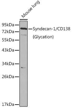
CD138 / Syndecan-1 Rabbit Polyclonal Antibody
The definitive plasma cell marker, used to identify them in tissue and marrow.
View product →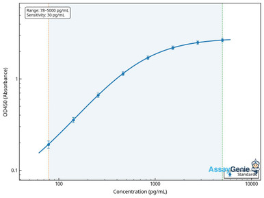
Human CD22 ELISA Kit
An inhibitory co-receptor restricted to the B lineage, measurable in soluble form.
View product →What CD markers are
CD stands for cluster of differentiation — a naming system agreed internationally so that a surface molecule recognised by several different antibodies gets one number rather than several competing names. The number reflects the order of assignment, not function or relatedness.
For B cells the practical value is that the marker profile changes predictably with developmental stage and activation state, so a panel of four or five antibodies can place a cell precisely in the lineage.
The core B cell markers
| Marker | Expression window | Why it matters |
|---|---|---|
| CD19 | Pro-B through plasma cell (reduced but present) | The most dependable pan-B marker across the whole lineage |
| CD20 | Pre-B through memory; absent on plasma cells | Target of anti-CD20 therapy — which is why such therapy does not remove existing plasma cells |
| CD21 | Mature B cells; high on marginal zone | Complement receptor 2; forms the CD19/CD21/CD81 co-receptor complex with the BCR |
| CD22 | B lineage restricted | Inhibitory co-receptor raising the BCR signalling threshold |
| CD27 | Memory B cells and plasma cells | The canonical human memory marker |
| CD38 | Germinal centre, transitional, plasma cells | High on both the earliest and latest stages, so never interpret it alone |
| CD138 | Plasma cells | Syndecan-1; the definitive plasma cell marker |
Why CD19 and CD20 are not interchangeable
Both are described as pan-B markers, but their windows differ at the ends of the lineage and the difference is clinically decisive.
CD19 appears early, is retained through differentiation, and remains detectable at lower density on plasma cells. CD20 appears somewhat later and is lost as cells become plasma cells.
The consequence: anti-CD20 depletion removes naive, memory and most mature B cells but leaves plasma cells untouched — so existing antibody titres persist. Anti-CD19 approaches, including CAR-T, reach further along the lineage. Choosing between them is a decision about which populations are meant to survive.
Markers through B cell development
| Stage | Characteristic markers | Defining event | |
|---|---|---|---|
| Progenitor / early pro-B | CD34, CD10, CD45, CD117 | Commitment to lymphoid lineage; CD34 marks progenitors and is lost thereafter | undefined |
| Pro-B | CD10, CD19, CD38, CD40, CD117 | Heavy chain D-J then V-DJ rearrangement begins | undefined |
| Pre-B | CD10, CD19, CD20 (low), CD40, CD93, CD127, CD25 | Pre-BCR expressed — rearranged heavy chain with surrogate light chain | undefined |
| Immature B | CD19, CD20, CD22, CD40, CD93; surface IgM positive | Light chain rearrangement complete — surface IgM is what defines this stage | undefined |
| Transitional B | CD19, CD10, CD21, CD24 (high), CD38 (high), CD93 | Migration from marrow to periphery; CD24/CD38 high is the defining profile | undefined |
| Naive mature B | CD19, CD20, CD21, CD40, CD185 (CXCR5), IgM and IgD positive | Full maturity, awaiting antigen | undefined |
Two corrections to values that circulate widely. CD34 belongs at the progenitor stage only and should not appear in an immature B phenotype. And CD28 is a T cell costimulatory receptor — it appears on plasma cells in some contexts, notably myeloma, but is not part of the immature B profile.
Most importantly, immature B cells are defined by surface IgM. Any description saying IgM is absent at this stage has the defining feature backwards.
Mature B cell subsets
| Subset | Marker profile | Note | |
|---|---|---|---|
| Follicular B | CD19+, CD20+, CD21 intermediate, CD23+, IgD+ | The conventional recirculating population; CD23 positivity is the discriminator | undefined |
| Marginal zone B | CD19+, CD21 high, CD23 low/negative, CD1d+, IgM high | Splenic, rapid-responding; the CD21-high CD23-negative profile separates it from follicular | undefined |
| Germinal centre B | CD19+, CD20+, CD38+, CD10+, CD95+, CD23 negative, CD27 negative, IgD negative | Site of somatic hypermutation and class switching | undefined |
| Memory B | CD19+, CD20+, CD27+, CD40+, often CD80+ | CD27 is the defining human memory marker | undefined |
| Plasmablast | CD19+, CD20 low/negative, CD27 high, CD38 high, CD138 low | Transitional, still proliferating | undefined |
| Plasma cell | CD19 low, CD20 negative, CD27+, CD38 high, CD138+, CD269 (BCMA)+ | Terminally differentiated and antibody-secreting | undefined |
The CD23 pattern is worth stating plainly because it is often listed incorrectly. CD23 is a follicular B cell marker. Marginal zone and germinal centre B cells are CD23-low or negative, and that is precisely how they are separated from the follicular population.
Likewise, memory B cells without CD27 in the panel cannot be reliably distinguished from naive cells — CD20 and CD40 alone do not do it, since both are present on naive B cells too.
B1 and B2 cells
The numbering here is counterintuitive and frequently reversed, so it is worth being explicit.
| B1 cells | B2 cells | |
|---|---|---|
| Which is conventional? | No — innate-like | Yes — the conventional follicular population |
| Location | Peritoneal and pleural cavities | Lymphoid follicles; recirculating |
| Antibody | Natural, polyreactive, mostly IgM | High-affinity, class-switched after antigen |
| Renewal | Self-renewing in the periphery | Continuously replaced from bone marrow |
So B2 is the conventional subset, not B1. The reverse assignment appears often enough that it is worth checking whenever the terms are used.
A gating strategy
| Step | Plot | Purpose |
|---|---|---|
| 1 | FSC-A vs SSC-A | Select lymphocytes, exclude debris |
| 2 | FSC-H vs FSC-A | Exclude doublets before anything else |
| 3 | Viability dye | Remove dead cells, which bind antibody non-specifically |
| 4 | CD19 vs CD3 | Identify B cells and exclude T cells in one step |
| 5 | CD27 vs IgD | Within CD19+: naive (IgD+CD27−), memory (CD27+), switched memory (IgD−CD27+) |
| 6 | CD38 vs CD24 | Resolve transitional cells, both high |
Including CD3 in the same plot as CD19 is a small but useful habit — it confirms the B gate is clean rather than merely assuming it.
Clonality and light chain restriction
A point absent from most marker lists but central to why B cell immunophenotyping is done clinically at all.
Each B cell expresses either a kappa or a lambda light chain, never both. A normal polyclonal population shows a kappa:lambda ratio of roughly 2:1. A markedly skewed ratio, or a population expressing only one light chain, indicates clonality and therefore a likely neoplastic process.
No surface marker identifies a malignant B cell directly. Light chain restriction is the readout that distinguishes an expanded reactive population from a clonal one, which is why anti-kappa and anti-lambda are standard in diagnostic B cell panels.
Clinical immunophenotypes
| Entity | Characteristic profile |
|---|---|
| Chronic lymphocytic leukaemia | CD19+, CD5+, CD23+, CD20 dim, surface immunoglobulin dim — the CD5/CD23 combination on a B cell is the signature |
| Mantle cell lymphoma | CD19+, CD5+, CD23 negative, cyclin D1+ — CD23 is what separates it from CLL |
| Follicular lymphoma | CD19+, CD10+, BCL2+, CD5 negative |
| Multiple myeloma | CD138+, CD38 high, CD19 negative, CD56 often positive — loss of CD19 distinguishes it from normal plasma cells |
| Precursor B-ALL | CD19+, CD10+, TdT+, CD34+, surface immunoglobulin negative |
Research and educational context only — diagnostic interpretation belongs with a qualified pathologist.
Controls
- FMO controls for each marker, which set gates correctly by accounting for spillover from every other channel in the panel.
- Viability dye, since dead cells bind antibody non-specifically and generate false positives.
- Single-stained compensation controls, using the same fluorophores as the panel.
- A known-positive sample — peripheral blood contains all the major B subsets and makes a convenient reference.
- Fc receptor blocking where myeloid cells are present, as their Fc receptors bind antibody irrespective of specificity.
Choosing antibodies
CD19 and CD20 for identification, CD27 for memory, CD38 and CD138 for plasma cells, and CD22 for B-lineage-restricted signalling work.
Browse B cell markers →Frequently asked questions
What is the difference between CD19 and CD20?
CD19 appears earlier and persists at reduced level on plasma cells; CD20 appears later and is absent from them. That is why anti-CD20 therapy depletes most B cells but leaves plasma cells and existing antibody titres intact.
Which marker identifies memory B cells?
CD27. It is the canonical human memory B cell marker, usually read against IgD to separate unswitched from class-switched memory. A memory panel without CD27 cannot reliably distinguish memory from naive cells.
Do immature B cells express surface IgM?
Yes — it is what defines the stage. Completion of light chain rearrangement produces surface IgM, and that is the transition from pre-B to immature B. Descriptions stating IgM is absent have the defining feature reversed.
Is CD23 a marginal zone B cell marker?
No. CD23 marks follicular B cells. Marginal zone and germinal centre B cells are CD23-low or negative, and that difference is exactly how follicular and marginal zone populations are told apart.
Are B1 cells the conventional B cells?
No — B2 cells are conventional. B1 cells are the innate-like, self-renewing population found mainly in the peritoneal and pleural cavities, producing natural polyreactive IgM. The numbering runs against intuition and is often stated backwards.
How is clonality assessed?
By kappa to lambda light chain ratio. Each B cell expresses one or the other, and a normal polyclonal population sits near 2:1. Marked skewing or restriction to a single light chain indicates a clonal population.
Which markers distinguish plasma cells?
CD138 with high CD38, CD27 positive, CD20 negative and CD19 reduced. In myeloma CD19 is typically lost altogether and CD56 is often gained, which separates malignant from normal plasma cells.
Recent Posts
-
Suspected Plague Death at a Siberian Anti-Plague Institute: What Researchers Should Know
Infectious Disease · News AnalysisSuspected Plague Death at a Siberian Anti-Plague Institute: What …6th Oct 2026 -
LPS ELISA Troubleshooting: The Importance of Sample Nature
Quick answer A researcher measuring circulating lipopolysaccharide (LPS) in human serum …25th Sep 2026 -

Sekundärantikörper wählen: Die Regel, die im Labor wirklich zählt
Sekundärantikörper wählen: Die Regel, die im Labor wirklich zählt Wählen Sie einen Sekundärantikörp …24th Sep 2026