
Platelets : The Unassuming Heroes of Blood Circulation
Platelets: Formation, Structure, Activation and Clinical Significance
Platelets are anucleate fragments shed from megakaryocytes, and they carry out primary haemostasis — adhering to damaged vessel wall, activating, and aggregating into a plug within seconds. This guide covers thrombopoiesis, the four structural zones, the receptors that mediate adhesion and aggregation, how activation is amplified, and the disorders and antiplatelet drugs that follow from each step.
Browse platelet assays →Key takeaways
- Platelets are anucleate cytoplasmic fragments released from megakaryocyte proplatelets — not cells in the conventional sense — and survive 7 to 10 days in circulation.
- Adhesion at high shear depends on von Willebrand factor bridging collagen to GPIb-IX-V; the direct collagen receptors are GPVI and integrin alpha-2-beta-1.
- Aggregation is mediated by GPIIb/IIIa binding fibrinogen, which is why its deficiency causes Glanzmann thrombasthenia and why it is a drug target.
- Thromboxane A2 is synthesised on demand from arachidonic acid via COX-1 — it is not stored in granules. That is precisely why aspirin, a COX-1 inhibitor, works as an antiplatelet agent.
- Dense granules store ADP, ATP, serotonin and calcium; alpha granules store proteins including von Willebrand factor, fibrinogen, PF4 and growth factors.
- Prostacyclin from healthy endothelium inhibits platelets, opposing thromboxane A2 — the two are the key antagonistic pair, alongside nitric oxide.
- Normal count is 150 to 450 × 109/L; below that is thrombocytopenia, above it thrombocytosis.
Contents
- What are platelets?
- Thrombopoiesis: how platelets form
- Shape and structure
- Adhesion, activation and aggregation
- Granule contents
- Amplification and the thromboxane pathway
- Platelets and immunity
- Platelet counts and their interpretation
- Platelet disorders
- Antiplatelet drugs
- Platelet transfusion
- Choosing platelet assays
- Frequently asked questions
Platelet ELISA kits
The panel follows the platelet lifecycle — production, adhesion, activation, granule release, eicosanoid output and the aggregation receptor itself.
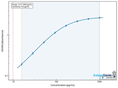
Human Thrombopoietin (THPO) ELISA Kit
The hormone driving megakaryocyte maturation and platelet production.
View product →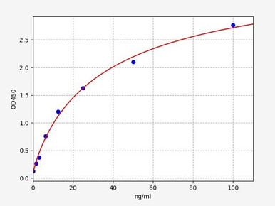
Human von Willebrand Factor ELISA Kit
Bridges collagen to GPIb-IX-V under high shear; deficiency causes the commonest inherited bleeding disorder.
View product →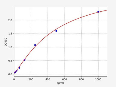
Human Soluble P-Selectin ELISA Kit
Surface-expressed on alpha granule release — the standard plasma marker of in vivo platelet activation.
View product →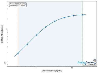
Human Platelet Factor 4 (PF4) ELISA Kit
An abundant alpha granule protein; central to heparin-induced thrombocytopenia.
View product →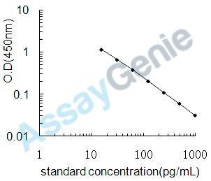
Thromboxane B2 (TXB2) ELISA Kit
TxA2 is too unstable to measure directly, so its stable metabolite TxB2 is quantified instead.
View product →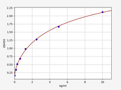
Human CD41 / Integrin alpha-IIb ELISA Kit
The alpha subunit of GPIIb/IIIa — the fibrinogen receptor deficient in Glanzmann thrombasthenia.
View product →What are platelets?
Platelets, also called thrombocytes, are small discoid structures circulating in blood at 150 to 450 × 109 per litre. They are central to haemostasis, the process that arrests bleeding after vascular injury.
A point of precision worth making at the outset: platelets are not cells. They are anucleate cytoplasmic fragments shed from megakaryocytes, lacking a nucleus and therefore unable to divide or mount a full transcriptional response. They do retain mitochondria and residual messenger RNA, so limited protein synthesis is possible, but the absence of a nucleus is what defines their biology and their finite 7 to 10 day lifespan.
The body produces on the order of 1011 platelets per day to maintain that count, and senescent platelets are cleared largely by the spleen and liver.
Thrombopoiesis: how platelets form
- Megakaryocyte development. Haematopoietic stem cells give rise to megakaryocyte-erythroid progenitors and then to the megakaryocyte lineage.
- Endomitosis. Maturing megakaryocytes replicate their DNA without dividing, becoming large and polyploid — commonly 16N to 64N. This unusual step provides the biosynthetic capacity to build thousands of platelets from one cell.
- Proplatelet formation. Mature megakaryocytes extend long proplatelet processes through the bone marrow sinusoidal endothelium into the bloodstream, where shear forces fragment them into individual platelets.
- Regulation. Thrombopoietin, produced constitutively by the liver, drives megakaryocyte proliferation and maturation. Its regulation is elegant: circulating platelets bind and clear TPO, so when platelet numbers fall, less TPO is removed, free TPO rises and production increases automatically.
A single megakaryocyte yields roughly 1,000 to 3,000 platelets. That autoregulatory TPO mechanism is also the rationale for TPO receptor agonists such as romiplostim and eltrombopag in chronic immune thrombocytopenia.
Shape and structure
Discoid shape and shape change
Resting platelets are discoid, which gives a high surface-area-to-volume ratio and allows them to travel at the vessel margin without obstructing flow. On activation they change shape dramatically, extending filopodia and lamellipodia that let them spread across damaged surfaces and interlock with one another. This shape change is cytoskeleton-driven and is one of the earliest visible signs of activation.
The four zones
| Zone | Contents | Function |
|---|---|---|
| Peripheral | Plasma membrane rich in glycoproteins and receptors | Adhesion and aggregation; senses the injured vessel wall |
| Sol-gel | Microtubule coil, actin and myosin | Maintains the discoid shape; drives shape change and clot retraction |
| Organelle | Alpha granules, dense granules, lysosomes, mitochondria | Stores and releases the mediators that amplify the response |
| Membrane systems | Open canalicular system and dense tubular system | The OCS provides a channel for rapid secretion and surface expansion; the DTS sequesters calcium |
The open canalicular system is worth noting as an elegant solution to a size problem: by invaginating the membrane throughout the interior, it massively increases available surface area and gives granules a route to release contents without each having to reach the outer membrane.
Adhesion, activation and aggregation
Primary haemostasis proceeds in three steps, each mediated by distinct receptors.
1. Adhesion
Vessel injury exposes subendothelial collagen. Under the high shear conditions of arterial flow, platelets cannot bind collagen directly at first — von Willebrand factor binds the exposed collagen, changes conformation under shear, and is then captured by the platelet receptor GPIb-IX-V. This tethering slows the platelet enough for firmer contacts to form.
The direct collagen receptors are GPVI, which delivers the main activating signal, and integrin alpha-2-beta-1, which provides stable adhesion. This distinction explains the two bleeding disorders: loss of GPIb-IX-V causes Bernard-Soulier syndrome, because the initial vWF tether fails.
2. Activation
Adhesion triggers intracellular signalling, raising cytosolic calcium and causing shape change, granule secretion and integrin activation. Thrombin generated by the coagulation cascade is the most potent physiological agonist, acting through protease-activated receptors.
3. Aggregation
Activation converts GPIIb/IIIa (integrin alpha-IIb-beta-3) from a low- to a high-affinity state — inside-out signalling. Activated GPIIb/IIIa binds fibrinogen, and because fibrinogen is bivalent it cross-links adjacent platelets into an aggregate. This is the final common step of aggregation regardless of the initiating agonist, which is why GPIIb/IIIa deficiency abolishes aggregation entirely in Glanzmann thrombasthenia.
The resulting platelet plug is then stabilised by fibrin from the coagulation cascade — secondary haemostasis — and activated platelets provide the phospholipid surface that cascade requires, so the two processes are interdependent rather than sequential.
Granule contents
| Granule | Contents | Role |
|---|---|---|
| Alpha granules | von Willebrand factor, fibrinogen, factor V, PF4, P-selectin, PDGF, TGF-beta, VEGF | The most numerous granules; supply adhesive proteins, coagulation factors and growth factors for repair |
| Dense granules | ADP, ATP, serotonin, calcium, polyphosphate | Small-molecule agonists that recruit and activate further platelets |
| Lysosomes | Acid hydrolases | Degrade matrix components and contribute to clot remodelling |
P-selectin is stored in alpha granule membranes and appears on the platelet surface only after degranulation, which is why surface or soluble P-selectin is used as a marker of activation rather than of platelet number.
Note what is not in the granules: thromboxane A2 is not stored anywhere in the platelet, as explained below.
Amplification and the thromboxane pathway
A few adherent platelets are not enough to stop bleeding. Activation is amplified by released mediators that recruit and activate passing platelets, producing a self-reinforcing response.
Thromboxane A2 is made, not stored
This point is frequently stated incorrectly, and the error matters clinically. Thromboxane A2 is not held in granules. On activation, phospholipase A2 liberates arachidonic acid from membrane phospholipid; cyclooxygenase-1 (COX-1) converts it to prostaglandin H2, and thromboxane synthase converts that to TxA2, which diffuses out to act on neighbouring platelets.
Because TxA2 is synthesised on demand by COX-1 rather than pre-packaged, irreversibly inhibiting COX-1 abolishes production for the platelet’s entire lifespan — it cannot transcribe replacement enzyme. That is exactly how low-dose aspirin works, and why its antiplatelet effect persists for days after the drug has cleared.
The amplification loop
- ADP released from dense granules acts on P2Y1 and P2Y12 receptors, sustaining aggregation. P2Y12 is the target of clopidogrel, prasugrel and ticagrelor.
- Thromboxane A2 newly synthesised acts on TP receptors, promoting aggregation and vasoconstriction.
- Thrombin from the coagulation cascade activates PAR1 and PAR4, the most potent stimulus of all.
- Calcium influx integrates these signals, driving shape change, secretion and integrin activation.
The endothelial brake
Healthy endothelium actively suppresses this. It releases prostacyclin (PGI2) and nitric oxide, both of which raise platelet cyclic nucleotides and inhibit activation, and it expresses CD39, which degrades ADP. So the balance is not simply thromboxane versus nitric oxide: prostacyclin and thromboxane are both prostanoids from the same arachidonic acid precursor, acting in opposite directions. Describing prostaglandins generally as pro-aggregatory is misleading, since the principal endothelial prostaglandin is inhibitory.
Platelets and immunity
Platelets are increasingly recognised as immune effectors rather than purely haemostatic fragments.
- Pathogen interaction. They express Toll-like receptors and can bind bacteria and viruses directly, sequestering them for clearance.
- Leukocyte recruitment. Surface P-selectin binds PSGL-1 on leukocytes, forming platelet-leukocyte aggregates that help neutrophils and monocytes reach injured tissue.
- Mediator release. Alpha granules contain chemokines including PF4 and RANTES, which recruit and activate immune cells.
- NET interaction. Platelets stimulate neutrophils to release extracellular traps, linking thrombosis and inflammation — the process termed immunothrombosis.
Platelet counts and their interpretation
| Count (×109/L) | Term | Significance |
|---|---|---|
| 150–450 | Normal | Adequate for haemostasis |
| 100–150 | Mild thrombocytopenia | Usually asymptomatic; investigate the cause |
| 50–100 | Moderate thrombocytopenia | Bleeding with trauma or surgery |
| 20–50 | Severe | Easy bruising, mucosal bleeding |
| Below 20 | Very severe | Risk of spontaneous bleeding |
| Above 450 | Thrombocytosis | Reactive in most cases; consider a myeloproliferative cause if persistent |
Counts are reported as ×109/L in SI units, equivalent to the per-microlitre figures used in some laboratories — 150 × 109/L is the same as 150,000 per µL.
One artefact worth knowing: pseudothrombocytopenia, in which EDTA in the sample tube causes platelets to clump in vitro, so the analyser reports a spuriously low count in a patient who is entirely well. Repeating in citrate resolves it, and it should be excluded before investigating an unexpected isolated low count.
Platelet disorders
Thrombocytopenia
Low counts arise through three mechanisms: reduced production (marrow failure, aplastic anaemia, leukaemia, chemotherapy), increased destruction (immune thrombocytopenia, drug-induced), or sequestration (splenomegaly). Presentation is mucocutaneous — petechiae, purpura, epistaxis — rather than the deep joint and muscle bleeding of coagulation factor deficiency.
Heparin-induced thrombocytopenia deserves separate mention: antibodies against PF4-heparin complexes activate platelets, so the count falls while the patient becomes prothrombotic rather than bleeding. It is the counterintuitive case, and it is why PF4 antibody testing exists.
Thrombocytosis
Reactive (secondary) thrombocytosis is far commoner, following inflammation, infection, iron deficiency, malignancy or surgery, and rarely causes thrombosis. Essential thrombocythaemia is a myeloproliferative neoplasm, frequently driven by JAK2, CALR or MPL mutations, and does carry thrombotic and haemorrhagic risk.
Function disorders
| Disorder | Defect | Consequence |
|---|---|---|
| Bernard-Soulier syndrome | GPIb-IX-V deficiency | Failure of vWF-mediated adhesion; large platelets and low count |
| Glanzmann thrombasthenia | GPIIb/IIIa deficiency | Aggregation fails entirely; count and platelet size normal |
| Gray platelet syndrome | Absent alpha granules | Grey appearance on film; mild to moderate bleeding, marrow fibrosis |
| Storage pool disease | Deficient dense granules | Impaired amplification through reduced ADP release |
| May-Hegglin anomaly | MYH9 mutation | Large platelets with Döhle-like leukocyte inclusions |
| von Willebrand disease | vWF deficiency or dysfunction | Commonest inherited bleeding disorder; platelets themselves are normal |
Antiplatelet drugs
Each major drug class maps onto a step described above, which is the clearest demonstration of why the mechanism matters.
| Class | Target | Examples |
|---|---|---|
| COX-1 inhibitors | Blocks thromboxane A2 synthesis irreversibly for the platelet lifespan | Low-dose aspirin |
| P2Y12 antagonists | Blocks the ADP amplification loop | Clopidogrel, prasugrel, ticagrelor |
| GPIIb/IIIa inhibitors | Blocks the final common aggregation step | Abciximab, eptifibatide, tirofiban |
| PAR1 antagonists | Blocks thrombin-mediated activation | Vorapaxar |
| Phosphodiesterase inhibitors | Raises platelet cyclic nucleotides, mimicking the endothelial brake | Dipyridamole, cilostazol |
Because platelets cannot synthesise new COX-1, aspirin’s effect lasts until the affected platelets are replaced — roughly 7 to 10 days — which is why it is stopped well before surgery. This is clinical context, not clinical advice.
Platelet transfusion
Platelet concentrates are prepared either by apheresis from a single donor or by pooling buffy coats from several whole blood donations. They are indicated for severe thrombocytopenia, for bleeding with platelet dysfunction, and prophylactically in haematology and oncology patients.
Storage constraints are unusual: platelets must be kept at room temperature with continuous agitation, because refrigeration activates them and shortens survival. That requirement limits shelf life to about five to seven days and carries a higher bacterial contamination risk than refrigerated components.
Risks include febrile and allergic reactions, transfusion-related acute lung injury, bacterial contamination, and alloimmunisation leading to refractoriness — where subsequent transfusions produce little increment. Decisions on transfusion belong with the treating clinician.
Choosing platelet assays
ELISA kits for thrombopoietin, von Willebrand factor, soluble P-selectin, platelet factor 4, thromboxane B2 and CD41 — covering production, adhesion, activation and granule release.
Browse platelet assays →Frequently asked questions
Are platelets cells?
Not in the conventional sense. They are anucleate cytoplasmic fragments shed from megakaryocytes. They retain mitochondria and some messenger RNA, allowing limited protein synthesis, but without a nucleus they cannot divide or mount a full transcriptional response.
Is thromboxane A2 stored in platelet granules?
No. It is synthesised on demand from membrane arachidonic acid by COX-1 and thromboxane synthase. Because platelets cannot make new COX-1, irreversible inhibition by aspirin lasts the whole platelet lifespan — which is the basis of its antiplatelet effect.
How do platelets stick to collagen?
Mostly indirectly at arterial shear. Von Willebrand factor binds exposed collagen, changes conformation under flow, and is captured by GPIb-IX-V on the platelet. The direct collagen receptors are GPVI, which signals activation, and integrin alpha-2-beta-1, which provides firm adhesion.
What is a normal platelet count?
150 to 450 × 109/L, equivalent to 150,000 to 450,000 per microlitre. Below 150 is thrombocytopenia and above 450 thrombocytosis, though an isolated low result should prompt exclusion of EDTA-induced pseudothrombocytopenia first.
How long do platelets live?
Seven to ten days in circulation. The body produces roughly 1011 platelets daily to maintain the count, and senescent platelets are cleared mainly by spleen and liver.
Why does heparin-induced thrombocytopenia cause clotting rather than bleeding?
Because the antibodies formed against PF4-heparin complexes activate platelets rather than simply destroying them. The count falls as platelets are consumed, but the patient becomes prothrombotic — the opposite of what a low count usually implies.
Why are platelets stored at room temperature?
Refrigeration activates platelets and causes rapid clearance after transfusion, so concentrates are kept at room temperature with continuous agitation. That constraint limits shelf life to about five to seven days and raises the bacterial contamination risk relative to refrigerated components.
Rithika Suresh completed her undergraduate degree in Biotechnology in Anna University before completing her masters in Biotechnology at University College Dublin.
Recent Posts
-
LPS ELISA Troubleshooting: The Importance of Sample Nature
Quick answer A researcher measuring circulating lipopolysaccharide (LPS) in human serum …25th Sep 2026 -

Sekundärantikörper wählen: Die Regel, die im Labor wirklich zählt
Sekundärantikörper wählen: Die Regel, die im Labor wirklich zählt Wählen Sie einen Sekundärantikörp …24th Sep 2026 -

Anticuerpos para IHC: cómo validarlos y evitar falsos positivos
Anticuerpos para IHC: cómo validarlos y evitar falsos positivos Use anticuerpos con evidencia publi …24th Sep 2026