Description
Custom Cell-Based ELISA Kit (BA0239) (BA0239)
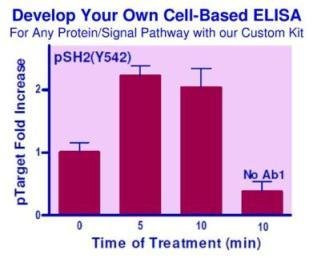
The Custom Cell-Based ELISA Kit (SKU: BA0239) is a flexible, rapid and sensitive starter tool for measuring the relative content of virtually any target protein directly in cultured cells. It requires only a target-specific mouse primary antibody (Ab1) supplied by the user, together with all the remaining reagents provided in the kit. The assay eliminates the need for cell lysate preparation: cells cultured in a 96-well plate are fixed and permeabilised, the target protein is detected using the user's Ab1 followed by a goat anti-mouse IgG-HRP (gM-HRP) secondary antibody and a fluorogenic substrate, and total protein is measured in the same well with a fluorescent protein stain. Target signal is normalised to total protein, greatly reducing well-to-well variation. It is well suited to studying protein content, signalling-pathway activity and the effects of drugs, inhibitors, siRNA or activators.
| Product Name: | Custom Cell-Based ELISA Kit (BA0239) |
| SKU: | BA0239 |
| Detection Method: | Cell-Based ELISA (FL530/585 nm, 360/450 nm) |
| Sample Type: | ['Cells'] |
| Species Reactivity: | Human, mouse, rat and more, depending on choice of primary antibody |
| Assay Time: | Assay takes 6.5 hrs; hands-on time 2.5 hrs |
| Kit Size: | 100 Assays |
| Equipment Required: | Microplate reader |
| Storage: | -20°C |
| Shelf Life: | 6 months |
| Shipping: | Gel Pack |
A do-it-yourself cell-based ELISA reagent kit that allows users to develop their own assay for any target protein for which a mouse primary antibody is available. The kit provides all components except the target-specific Ab1. Target protein is measured fluorimetrically at λex/em = 530/585 nm and normalised to total cellular protein measured at 360/450 nm in the same well.
- New and improved. Total assay time reduced from the standard 21 hours to 6.5 hours (hands-on time 2.5 hrs).
- Simple and convenient. Cells are directly cultured in 96-well plates. No cell lysis necessary.
- Accurate and high-throughput. Target protein is normalised to total cellular protein in the same well, greatly minimising well-to-well variations, and can be readily automated for thousands of samples per day.
- Determination of relative protein content and signal pathway (e.g. phosphorylation status) in whole cells.
- Evaluation of target modulation by drugs, activators, inhibitors, siRNA etc.
Note: The below protocol is a sample protocol. Protocols are specific to each batch/lot. For the correct instructions please follow the protocol included in your kit.
| Step | Procedure |
| 1 | Prepare 1x Wash Buffer by diluting Stock Wash Buffer 40-fold with dH2O (e.g. 10 mL Stock Wash Buffer + 390 mL dH2O). Reserve 6 mL for the Detection step. Assay samples in triplicate or more, and include a Protein Blank (no cells) and a Sample Blank (cells with no Ab1, only Ab2). |
| 2 | Culture and treat cells. Seed 100 µL of 1-3×10^4 adherent cells (or 4-10×10^4 suspension cells) per well of a black 96-well culture plate; add 100 µL cell-free media into three wells for the Protein Blank. Incubate overnight at 37°C, then treat cells as desired. |
| 3 | Fix cells (caution: formaldehyde is toxic; use a chemical hood and appropriate protection). For adherent cells, replace media with 100 µL 4% formaldehyde; for suspension cells, add 100 µL 8% formaldehyde to the pellet. Incubate 20 min at room temperature, then wash twice with 200 µL 1× Wash Buffer. |
| 4 | Quench. Add 100 µL Quench Buffer (2.2 mL 3% H2O2 + 8.8 mL 1× Wash Buffer) per well; incubate 20 min at room temperature, then wash three times with 200 µL 1× Wash Buffer. |
| 5 | Block. Add 100 µL Blocking Buffer per well; incubate 1 hr at room temperature. |
| 6 | Add Primary Antibody (Ab1). Perform a pilot titration (e.g. 1:500 - 1:5000) to determine the optimal Ab1 dilution. Add 50 µL Blocking Buffer to Sample Blank wells and 50 µL diluted Ab1 to Sample wells; incubate 90 min at room temperature (or overnight at 2-8°C) with gentle shaking, then wash three times. |
| 7 | Add Secondary Antibody (Ab2). Prepare Ab2 in Blocking Buffer at 1:1000; add 50 µL to all wells; incubate 90 min at room temperature with gentle shaking. |
| 8 | Detection. Wash four times with 200 µL 1× Wash Buffer. Prepare HRP Substrate (60 µL Dye Reagent + 6 mL 1× Wash Buffer + 6 µL 3% H2O2), add 50 µL per well and incubate 30 min at room temperature in the dark. Add 50 µL Protein Stain per well and incubate a further 5 min in the dark. |
| 9 | Read the plate at λex/em = 530/585 nm for target protein and at λex/em = 360/450 nm for total protein. |
Calculate the mean target fluorescence at 530/585 nm for the Sample Blank (“No Ab1” wells, F_Target.Blank) and Sample wells (F_Target.Sample), and the mean total protein fluorescence at 360/450 nm for the Protein Blank (F_Protein.Blank) and Sample wells (F_Protein.Sample). ΔF_Target = F_Target.Sample – F_Target.Blank; ΔF_Protein = F_Protein.Sample – F_Protein.Blank. Normalised Target = (ΔF_Target/ΔF_Protein) / (ΔF_Target/ΔF_Protein)O, where (ΔF_Target/ΔF_Protein)O is the control reference value (e.g. time zero or untreated wells).
| Component | Quantity | Storage |
| Stock Wash Buffer | 25 mL | -20°C |
| Blocking Buffer | 25 mL | -20°C |
| Protein Stain | 6 mL | -20°C |
| Dye Reagent | 120 µL | -20°C |
| Ab1 (mouse, target dependent) | not provided | -20°C |
| Ab2 (goat anti-mouse IgG-HRP (gM-HRP)) | 10 µL | -20°C |