The DISC1 Monoclonal Antibody (CAB4678) is a high-quality antibody developed for reliable detection and analysis of target proteins. This antibody, developed using rabbit monoclonal technology, offers high specificity and affinity for human samples, making it an ideal choice for research in neuroscience and mental health.DISC1, also known as disrupted in schizophrenia 1, is a crucial player in various cellular processes, including neuronal migration, neurite outgrowth, and synaptic transmission. Dysregulation of DISC1 has been linked to a range of psychiatric conditions, including schizophrenia, bipolar disorder, and major depressive disorder.
This antibody is validated for use in WB, IF/ICC, ELISA applications and has demonstrated reactivity against Human, Mouse, Rat samples.
Product Name:
DISC1 Monoclonal Antibody
SKU:
CAB4678
Size:
20μL, 100μL
Reactivity:
Human, Mouse, Rat
Clone Number:
ARC1089
Conjugate:
Unconjugated
Immunogen:
Synthetic peptide. This information is considered to be commercially sensitive.
This gene encodes a protein with multiple coiled coil motifs which is located in the nucleus, cytoplasm and mitochondria. The protein is involved in neurite outgrowth and cortical development through its interaction with other proteins. This gene is disrupted in a t(1;11)(q42.1;q14.3) translocation which segregates with schizophrenia and related psychiatric disorders in a large Scottish family. Alternate transcriptional splice variants, encoding different isoforms, have been characterized.
Purification Method
Affinity purification
Gene ID
27185
RRID
AB_2863321
Buffer Information
Store at -20℃. Avoid freeze / thaw cycles. Buffer: PBS containing 50% glycerol and 0.05% BSA, preserved with proclin300 or sodium azide, pH 7.3.
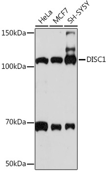
Western blot analysis of various lysates using DISC1 Rabbit mAb (CAB4678) at 1:1000 dilution. Secondary antibody: HRP-conjugated Goat anti-Rabbit IgG (H+L) (CABS014) at 1:10000 dilution. Lysates/proteins: 25μg per lane. Blocking buffer: 3% nonfat dry milk in TBST. Detection: ECL Basic Kit (AbGn00020). Exposure time: 10s.
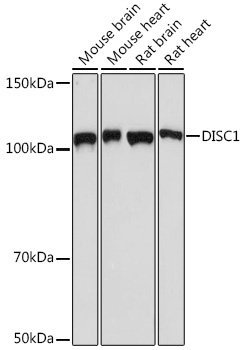
Western blot analysis of various lysates using DISC1 Rabbit mAb (CAB4678) at 1:1000 dilution. Secondary antibody: HRP-conjugated Goat anti-Rabbit IgG (H+L) (CABS014) at 1:10000 dilution. Lysates/proteins: 25μg per lane. Blocking buffer: 3% nonfat dry milk in TBST. Detection: ECL Basic Kit (AbGn00020). Exposure time: 30s.
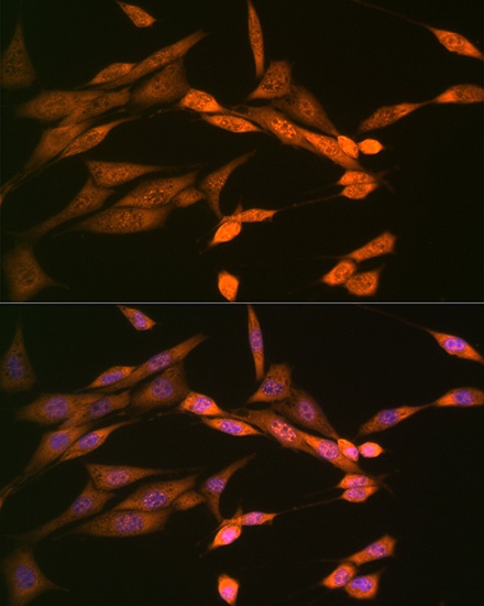
Immunofluorescence analysis of NIH-3T3 cells using DISC1 Rabbit mAb (CAB4678) at dilution of 1:100 (40x lens). Secondary antibody: Cy3-conjugated Goat anti-Rabbit IgG (H+L) (CABS007) at 1:500 dilution. Blue: DAPI for nuclear staining.
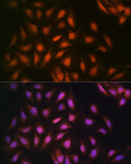
Immunofluorescence analysis of U-2 OS cells using DISC1 Rabbit mAb (CAB4678) at dilution of 1:100 (40x lens). Secondary antibody: Cy3-conjugated Goat anti-Rabbit IgG (H+L) (CABS007) at 1:500 dilution. Blue: DAPI for nuclear staining.


![Anti-DISC1 [R03-3M7] Monoclonal Antibody (AGMB03170)](https://cdn11.bigcommerce.com/s-h68l9z2lnx/images/stencil/590x590/products/274459/678031/anti-disc1-r03-3m7-monoclonal-antibody-agmb03170__18442.1773034468.jpg?c=2 "Anti-DISC1 [R03-3M7] Monoclonal Antibody (AGMB03170)")
![Anti-DISC1 [R03-7F5] Monoclonal Antibody (AGMB00199)](https://cdn11.bigcommerce.com/s-h68l9z2lnx/images/stencil/590x590/products/271488/691583/anti-disc1-r03-7f5-monoclonal-antibody-agmb00199__42144.1774503448.jpg?c=2 "Anti-DISC1 [R03-7F5] Monoclonal Antibody (AGMB00199)")


