The HES1 Monoclonal Antibody (CAB0925) is a high-quality antibody developed for reliable detection and analysis of target proteins. This antibody, produced through rabbit monoclonal technology, is well-suited for use in various applications, including immunofluorescence, immunohistochemistry, and flow cytometry.HES1 is known to play a key role in regulating stem cell maintenance and differentiation, making it a crucial target for research in developmental biology and cancer biology. The HES1 Rabbit Monoclonal Antibody allows for precise detection and visualization of HES1 protein expression in different cell types and tissues, providing valuable insights into its function and mechanisms of action.
This antibody is validated for use in WB, IHC-P, IP, ELISA applications and has demonstrated reactivity against Human, Mouse, Rat samples.
Product Name:
HES1 Monoclonal Antibody
SKU:
CAB0925
Size:
20μL, 100μL
Reactivity:
Human, Mouse, Rat
Clone Number:
ARC0513
Conjugate:
Unconjugated
Immunogen:
Synthetic peptide. This information is considered to be commercially sensitive.
0.5μg-4μg antibody for 400μg-600μg extracts of whole cells
ELISA
Recommended starting concentration is 1 μg/mL. Please optimize the concentration based on your specific assay requirements.
Synonyms:
HHL, HRY, HES-1, bHLHb39, HES1
Positive Sample:
MOLT-4, Mouse testis
Cellular Localization:
Nucleus.
Calculated MW:
30kDa
Observed MW:
30kDa
This protein belongs to the basic helix-loop-helix family of transcription factors. It is a transcriptional repressor of genes that require a bHLH protein for their transcription. The protein has a particular type of basic domain that contains a helix interrupting protein that binds to the N-box rather than the canonical E-box.
Purification Method
Affinity purification
Gene ID
3280
RRID
AB_2861486
Buffer Information
Store at -20℃. Avoid freeze / thaw cycles. Buffer: PBS containing 50% glycerol and 0.05% BSA, preserved with proclin300 or sodium azide, pH 7.3.
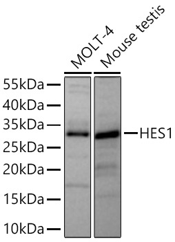
Western blot analysis of various lysates using HES1 Rabbit mAb (CAB0925) at 1:1000 dilution incubated overnight at 4℃. Secondary antibody: HRP-conjugated Goat anti-Rabbit IgG (H+L) (CABS014) at 1:10000 dilution. Lysates/proteins: 25 μg per lane. Blocking buffer: 3% nonfat dry milk in TBST. Detection: ECL Basic Kit (AbGn00020). Exposure time: 45s.
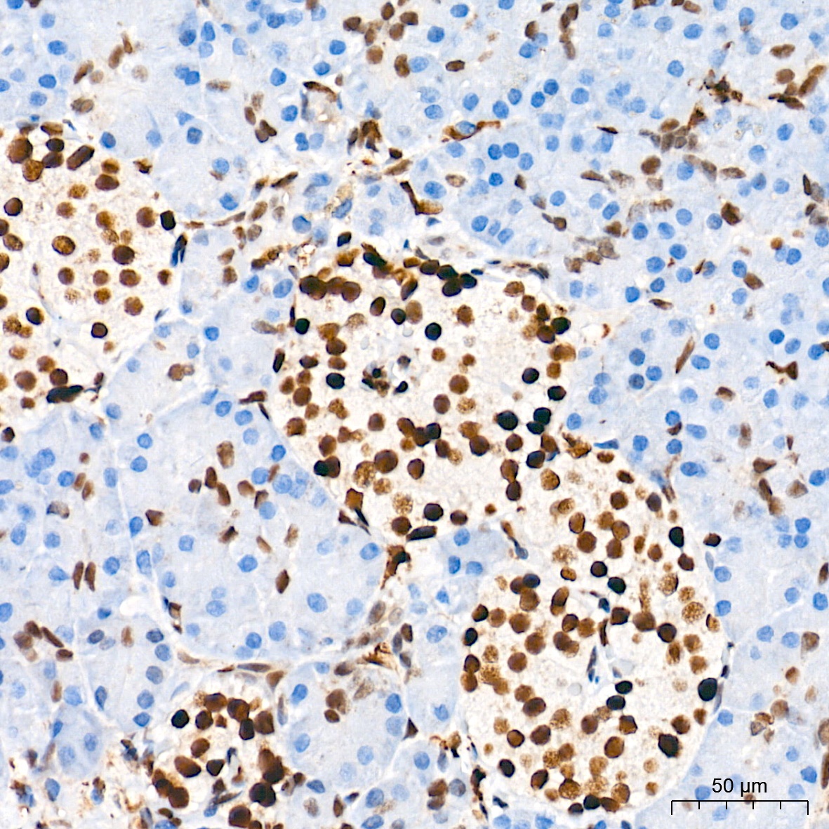
Immunohistochemistry analysis of paraffin-embedded Human pancreas tissue using HES1 Rabbit mAb (CAB0925) at a dilution of 1:2000 (40x lens). High pressure antigen retrieval performed with 0.01M Tris-EDTA Buffer (pH 9.0) prior to IHC staining.
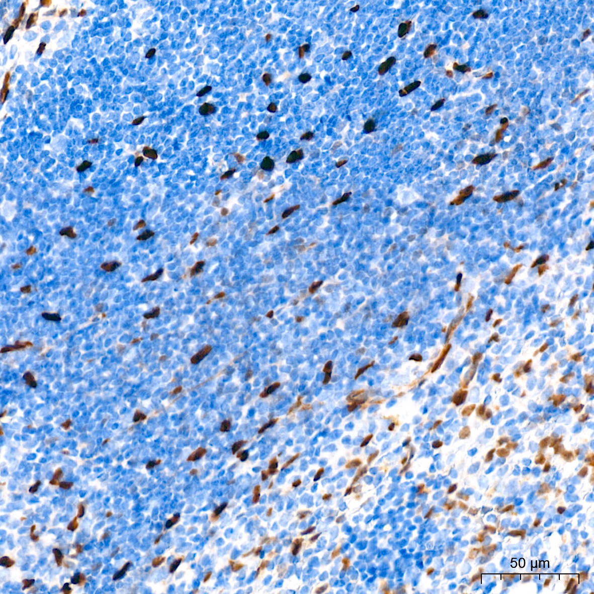
Immunohistochemistry analysis of paraffin-embedded Mouse spleen tissue using HES1 Rabbit mAb (CAB0925) at a dilution of 1:2000 (40x lens). High pressure antigen retrieval performed with 0.01M Tris-EDTA Buffer (pH 9.0) prior to IHC staining.
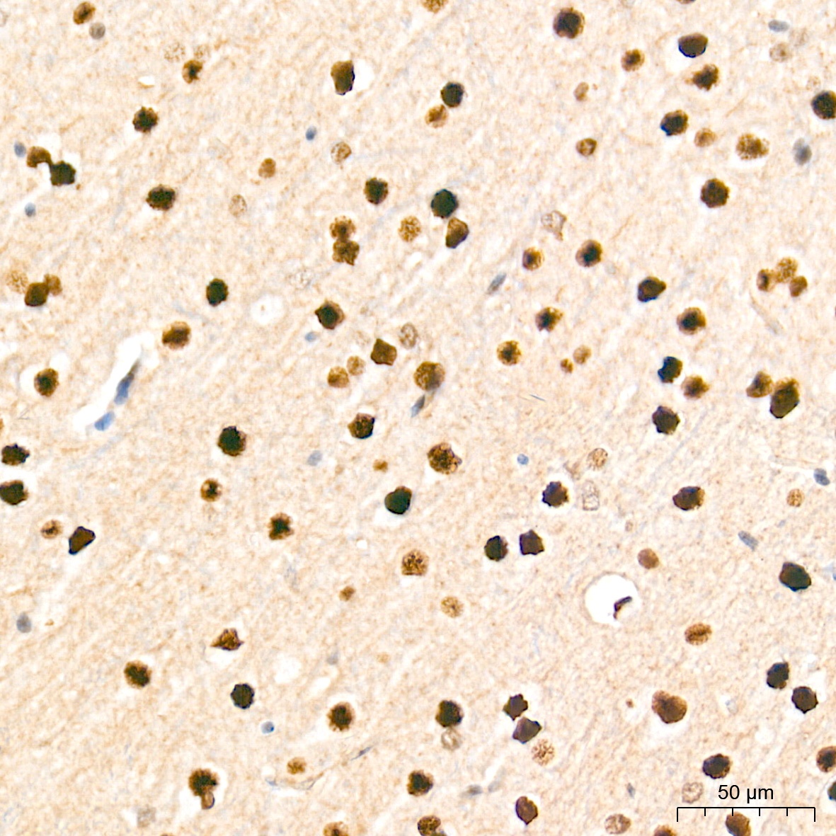
Immunohistochemistry analysis of paraffin-embedded Rat brain tissue using HES1 Rabbit mAb (CAB0925) at a dilution of 1:2000 (40x lens). High pressure antigen retrieval performed with 0.01M Tris-EDTA Buffer (pH 9.0) prior to IHC staining.
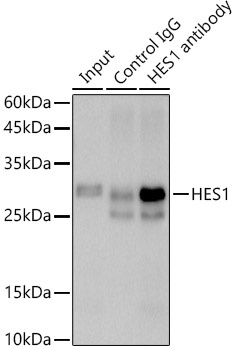
Immunoprecipitation analysis of 600 μg extracts of Rat testis cells using 3 μg HES1 antibody (CAB0925). Western blot was performed from the immunoprecipitate using HES1 antibody (CAB0925) at a dilution of 1:500.


![Anti-HES1 [R01-6B1] Monoclonal Antibody (AGMB00775)](https://cdn11.bigcommerce.com/s-h68l9z2lnx/images/stencil/590x590/products/272064/693576/anti-hes1-r01-6b1-monoclonal-antibody-agmb00775__98154.1774509788.jpg?c=2 "Anti-HES1 [R01-6B1] Monoclonal Antibody (AGMB00775)")


