The Phospho-p53-S33 Antibody (CABP0762) is a high-quality antibody developed for reliable detection and analysis of target proteins. This antibody, raised in rabbits, is highly specific for detecting phosphorylation of TP53 at serine 33 in human samples, and has been validated for use in Western blot applications.Phosphorylation of TP53 at serine 33 is known to regulate its activity and stability, influencing its ability to induce cell cycle arrest, apoptosis, and DNA repair in response to cellular stress. The Phospho-TP53 (S33) Polyclonal Antibody enables researchers to study the phosphorylation status of TP53 in various cell types, providing valuable insights into the mechanisms of TP53-mediated tumorigenesis and potential therapeutic strategies targeting this pathway.
This antibody is validated for use in WB, IHC-P, IP, ELISA applications and has demonstrated reactivity against Human, Mouse, Rat samples.
Product Name:
Phospho-p53-S33 Antibody
SKU:
CABP0762
Size:
20μL, 100μL
Reactivity:
Human, Mouse, Rat
Conjugate:
Unconjugated
Immunogen:
Synthetic peptide. This information is considered to be commercially sensitive.
Sequence:
VLSP L
Tested Applications:
WBIHC-PIPELISA
Recommended Dilution:
WB
1:500 - 1:2000
IHC-P
1:50 - 1:100
IP
0.5μg-4μg antibody for 200μg-400μg extracts of whole cells
ELISA
Recommended starting concentration is 1 μg/mL. Please optimize the concentration based on your specific assay requirements.
This gene encodes a tumor suppressor protein containing transcriptional activation, DNA binding, and oligomerization domains. The encoded protein responds to diverse cellular stresses to regulate expression of target genes, thereby inducing cell cycle arrest, apoptosis, senescence, DNA repair, or changes in metabolism. Mutations in this gene are associated with a variety of human cancers, including hereditary cancers such as Li-Fraumeni syndrome. Alternative splicing of this gene and the use of alternate promoters result in multiple transcript variants and isoforms. Additional isoforms have also been shown to result from the use of alternate translation initiation codons from identical transcript variants (PMIDs: 12032546, 20937277).
Purification Method
Affinity purification
Gene ID
7157
RRID
AB_2771619
Buffer Information
Store at -20℃. Avoid freeze / thaw cycles. Buffer: PBS with 0.01% thimerosal,50% glycerol,pH7.3.
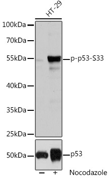
Western blot analysis of lysates from HT-29 cells, using Phospho-p53-S33 Rabbit pAb (CAB3185). HT-29 cells were treated with Nocodazole (100ng/mL) for 16 hours. Secondary antibody: HRP-conjugated Goat anti-Rabbit IgG (H+L) (CABS014) at 1:10000 dilution. Lysates/proteins: 25μg per lane. Blocking buffer: 3% BSA. Detection: ECL Basic Kit (AbGn00020). Exposure time: 1s.
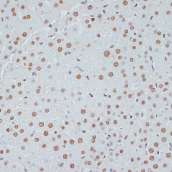
Immunohistochemistry analysis of paraffin-embedded Rat liver using Phospho-p53-S33 Rabbit pAb (CABP0762) at dilution of 1:100 (40x lens). Microwave antigen retrieval performed with 0.01M Tris/EDTA Buffer (pH 9.0) prior to IHC staining.
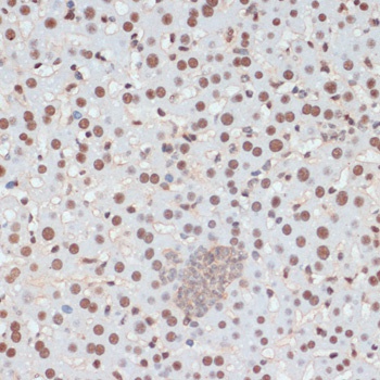
Immunohistochemistry analysis of paraffin-embedded Mouse liver using Phospho-p53-S33 Rabbit pAb (CABP0762) at dilution of 1:100 (40x lens). Microwave antigen retrieval performed with 0.01M Tris/EDTA Buffer (pH 9.0) prior to IHC staining.
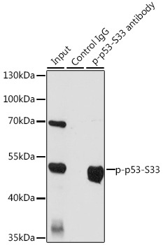
Immunoprecipitation analysis of 200 μg extracts of HT-29 cells, using 3 μg Phospho-p53-S33 pAb (CABP0762). Western blot was performed from the immunoprecipitate using Phospho-p53-S33 pAb (CABP0762) at a dilution of 1:1000. HT-29 cells were treated with nocodazole (100 ng/mL) at 37℃ for 16 hours.