The BCL2L14/BCLG Monoclonal Antibody (CAB19292) is a high-quality antibody developed for reliable detection and analysis of target proteins. This polyclonal antibody, generated in rabbits, is highly specific to human samples and has been validated for use in Western blotting applications.BCL2L14/BCLG is a member of the BCL-2 family of proteins, known for their roles in controlling cell death and survival pathways. Research has shown that BCL2L14/BCLG may play a role in modulating apoptosis and influencing cancer cell growth.
This antibody is validated for use in WB, IHC-P, ELISA applications and has demonstrated reactivity against Human, Mouse, Rat samples.
Product Name:
BCL2L14/BCLG Monoclonal Antibody
SKU:
CAB19292
Size:
20μL, 100μL
Reactivity:
Human, Mouse, Rat
Clone Number:
ARC2474
Conjugate:
Unconjugated
Immunogen:
Recombinant protein (or fragment).This information is considered to be commercially sensitive.
Recommended starting concentration is 1 μg/mL. Please optimize the concentration based on your specific assay requirements.
Synonyms:
BCLG, BCL2L14/BCLG
Positive Sample:
A549, BxPC-3, Mouse large intestine, Mouse testis, Mouse stomach, Rat testis
Cellular Localization:
Cytoplasm, Endomembrane System, Cytosol.
Calculated MW:
37kDa
Observed MW:
37kDa
The protein encoded by this gene belongs to the BCL2 protein family. BCL2 family members form hetero- or homodimers and act as anti- or pro-apoptotic regulators that are involved in a wide variety of cellular activities. Overexpression of this gene has been shown to induce apoptosis in cells. Three alternatively spliced transcript variants encoding two distinct isoforms have been reported for this gene.
Purification Method
Affinity purification
Gene ID
79370
Buffer Information
Store at -20℃. Avoid freeze / thaw cycles. Buffer: PBS containing 50% glycerol and 0.05% BSA, preserved with proclin300 or sodium azide, pH 7.3.
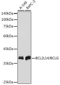
Western blot analysis of various lysates using BCL2L14/BCLG Rabbit mAb (CAB19292) at 1:1000 dilution. Secondary antibody: HRP-conjugated Goat anti-Rabbit IgG (H+L) (CABS014) at 1:10000 dilution. Lysates/proteins: 25μg per lane. Blocking buffer: 3% nonfat dry milk in TBST. Detection: ECL Basic Kit (AbGn00020). Exposure time: 1s.
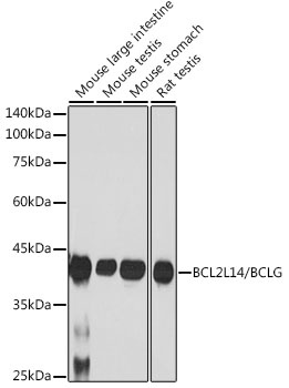
Western blot analysis of various lysates using BCL2L14/BCLG Rabbit mAb (CAB19292) at 1:1000 dilution. Secondary antibody: HRP-conjugated Goat anti-Rabbit IgG (H+L) (CABS014) at 1:10000 dilution. Lysates/proteins: 25μg per lane. Blocking buffer: 3% nonfat dry milk in TBST. Detection: ECL Basic Kit (AbGn00020). Exposure time: 180s.
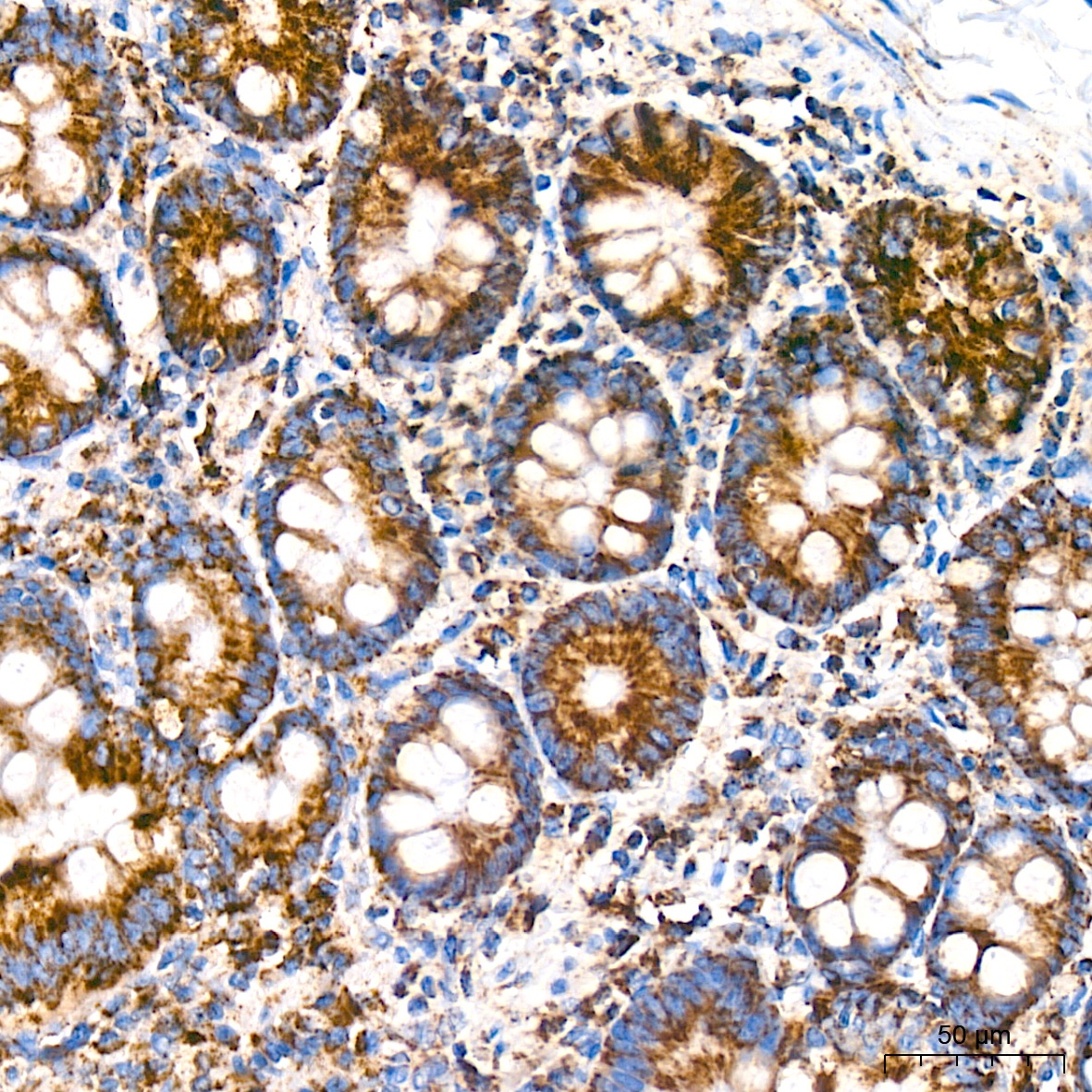
Immunohistochemistry analysis of paraffin-embedded Human colon tissue using BCL2L14/BCLG Rabbit mAb (CAB19292) at a dilution of 1:200 (40x lens). High pressure antigen retrieval was performed with 0.01 M Tris-EDTA buffer (pH 9.0) prior to IHC staining.
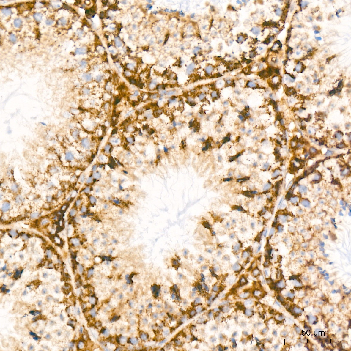
Immunohistochemistry analysis of paraffin-embedded Mouse testis tissue using BCL2L14/BCLG Rabbit mAb (CAB19292) at a dilution of 1:200 (40x lens). High pressure antigen retrieval was performed with 0.01 M Tris-EDTA buffer (pH 9.0) prior to IHC staining.
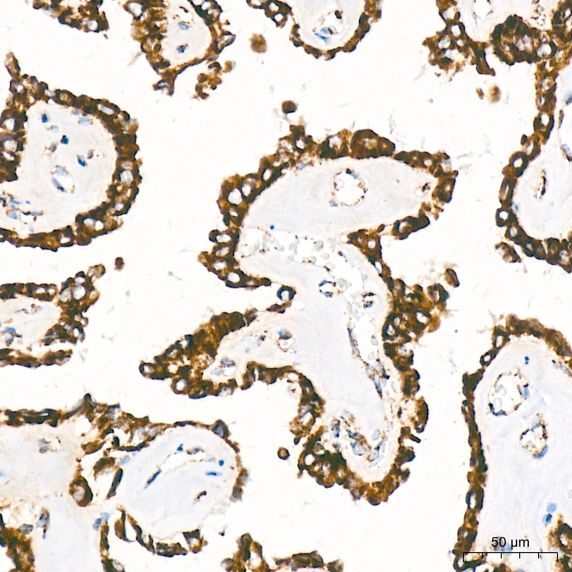
Immunohistochemistry analysis of paraffin-embedded Human lung adenocarcinoma tissue using BCL2L14/BCLG Rabbit mAb (CAB19292) at a dilution of 1:200 (40x lens). High pressure antigen retrieval was performed with 0.01 M Tris-EDTA buffer (pH 9.0) prior to IHC staining.
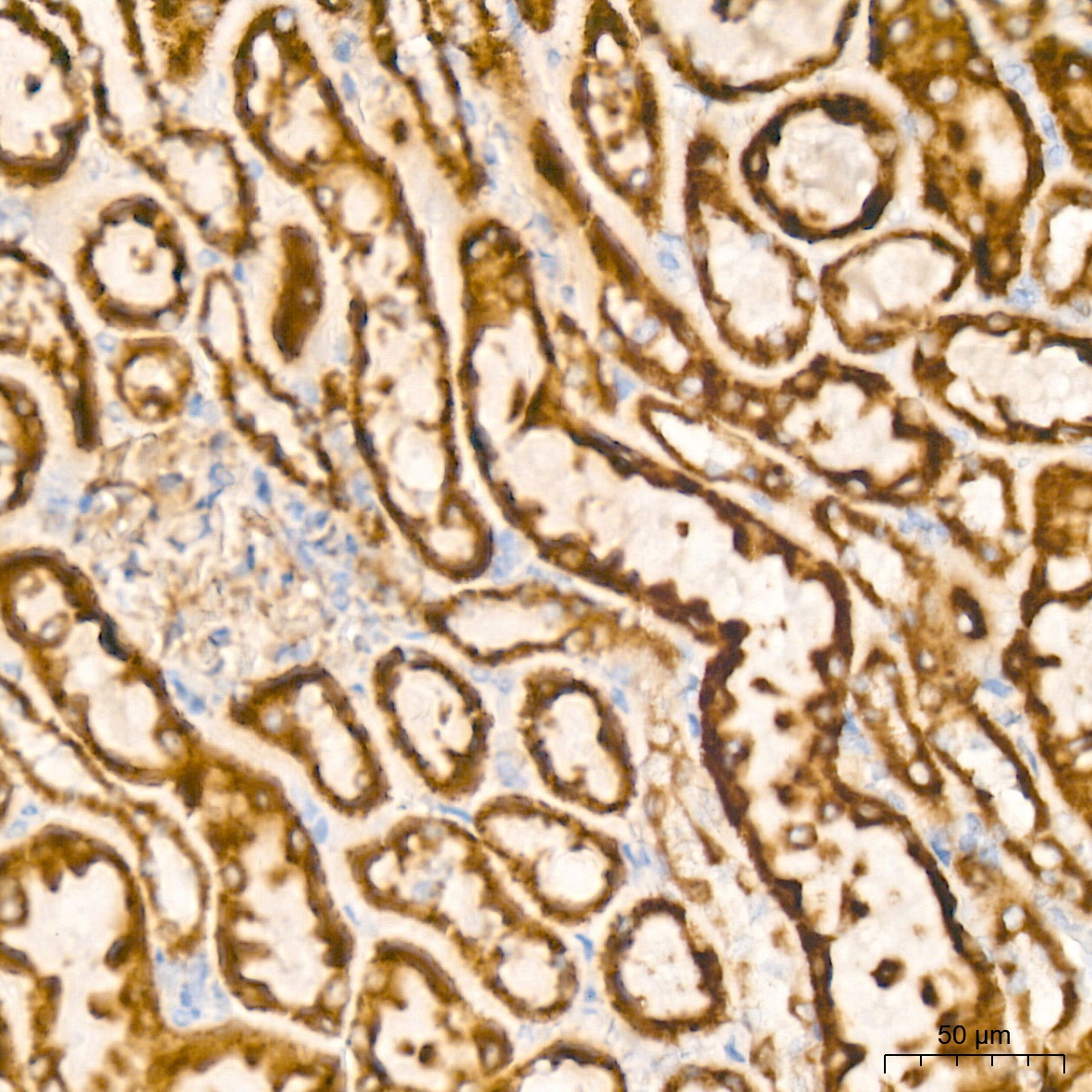
Immunohistochemistry analysis of paraffin-embedded Rat kidney tissue using BCL2L14/BCLG Rabbit mAb (CAB19292) at a dilution of 1:200 (40x lens). High pressure antigen retrieval was performed with 0.01 M Tris-EDTA buffer (pH 9.0) prior to IHC staining.


![Anti-BCL2L14/BCLG [R06-3D8] Monoclonal Antibody (AGMB00289)](https://cdn11.bigcommerce.com/s-h68l9z2lnx/images/stencil/590x590/products/271578/693243/anti-bcl2l14bclg-r06-3d8-monoclonal-antibody-agmb00289__22030.1774508718.jpg?c=2 "Anti-BCL2L14/BCLG [R06-3D8] Monoclonal Antibody (AGMB00289)")
![Anti-BCL2L14/BCLG [4X6-C9-E7] Monoclonal Antibody (AGMB04931)](https://cdn11.bigcommerce.com/s-h68l9z2lnx/images/stencil/590x590/products/276216/678223/anti-bcl2l14bclg-4x6-c9-e7-monoclonal-antibody-agmb04931__37371.1773035071.jpg?c=2 "Anti-BCL2L14/BCLG [4X6-C9-E7] Monoclonal Antibody (AGMB04931)")


