The ARID3A Antibody (CAB7668) is a high-quality antibody developed for reliable detection and analysis of target proteins. This antibody, produced through rabbit immunization, exhibits high reactivity with human samples and is validated for use in a variety of applications, including Western blotting.ARID3A, also known as Bright, is a transcription factor that plays a critical role in controlling gene expression and cellular development. Its involvement in regulating cell fate decisions makes it a crucial target for investigations in developmental biology, cancer research, and stem cell studies. The ARID3A Polyclonal Antibody enables precise detection and quantification of ARID3A protein levels in different cell types, facilitating detailed analysis and insights into its function.
This antibody is validated for use in WB, IHC-P, IP, ELISA applications and has demonstrated reactivity against Human, Mouse, Rat samples.
Product Name:
ARID3A Antibody
SKU:
CAB7668
Size:
20μL, 100μL
Reactivity:
Human, Mouse, Rat
Conjugate:
Unconjugated
Immunogen:
Recombinant protein (or fragment).This information is considered to be commercially sensitive.
0.5μg-4μg antibody for 200μg-400μg extracts of whole cells
ELISA
Recommended starting concentration is 1 μg/mL. Please optimize the concentration based on your specific assay requirements.
Synonyms:
DRIL1, DRIL3, BRIGHT, E2FBP1, ARID3A
Positive Sample:
K-562
Cellular Localization:
Cytoplasm, Nucleus.
Calculated MW:
63kDa
Observed MW:
75kDa
This gene encodes a member of the ARID (AT-rich interaction domain) family of DNA binding proteins. It was found by homology to the Drosophila dead ringer gene, which is important for normal embryogenesis. Other ARID family members have roles in embryonic patterning, cell lineage gene regulation, cell cycle control, transcriptional regulation, and possibly in chromatin structure modification.
Purification Method
Affinity purification
Gene ID
1820
RRID
AB_2768437
Buffer Information
Store at -20℃. Avoid freeze / thaw cycles. Buffer: PBS containing 50% glycerol, preserved with proclin300 or sodium azide, pH 7.3.
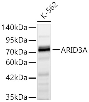
Western blot analysis of lysates from K-562 cells using ARID3A Rabbit pAb (CAB7668) at 1:1000 dilution. Secondary antibody: HRP-conjugated Goat anti-Rabbit IgG (H+L) (CABS014) at 1:10000 dilution. Lysates/proteins: 25 μg per lane. Blocking buffer: 3% nonfat dry milk in TBST. Detection: ECL Basic Kit (AbGn00020). Exposure time: 10s.
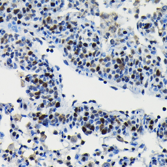
Immunohistochemistry analysis of paraffin-embedded Rat lung using ARID3A Rabbit pAb (CAB7668) at dilution of 1:100 (40x lens). High pressure antigen retrieval performed with 0.01M Citrate buffer (pH 6.0) prior to IHC staining.
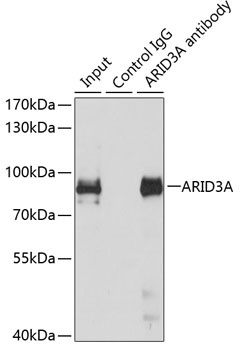
Immunoprecipitation analysis of 150 μg extracts of MCF7 cells using 3 μg ARID3A antibody (CAB7668). Western blot was performed from the immunoprecipitate using ARID3A antibody (CAB7668) at a dilution of 1:500.