The BNIP1 Antibody (CAB7263) is a high-quality antibody developed for reliable detection and analysis of target proteins. This antibody is produced in rabbits and exhibits high reactivity towards human samples, making it an excellent choice for Western blot applications. By binding specifically to BNIP1, this antibody allows for the detection and analysis of BNIP1 protein levels in a variety of cell types.BNIP1 is a key player in cellular processes such as programmed cell death and cellular energy metabolism, highlighting its importance in the field of cancer research and neurodegenerative diseases.
This antibody is validated for use in WB, ELISA applications and has demonstrated reactivity against Human, Mouse, Rat samples.
Product Name:
BNIP1 Antibody
SKU:
CAB7263
Size:
20μL, 100μL
Reactivity:
Human, Mouse, Rat
Conjugate:
Unconjugated
Immunogen:
Recombinant protein (or fragment).This information is considered to be commercially sensitive.
Recommended starting concentration is 1 μg/mL. Please optimize the concentration based on your specific assay requirements.
Synonyms:
NIP1, SEC20, TRG-8, BNIP1
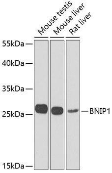
Positive Sample:
Mouse testis, Mouse liver, Rat liver
Cellular Localization:
Endoplasmic Reticulum Membrane, Mitochondrion, Single-Pass Type Iv Membrane Protein.
Calculated MW:
26kDa
Observed MW:
26kDa
This gene is a member of the BCL2/adenovirus E1B 19 kd-interacting protein (BNIP) family. It interacts with the E1B 19 kDa protein, which protects cells from virally-induced cell death. The encoded protein also interacts with E1B 19 kDa-like sequences of BCL2, another apoptotic protector. In addition, this protein is involved in vesicle transport into the endoplasmic reticulum. Alternative splicing of this gene results in four protein products with identical N- and C-termini.
Purification Method
Affinity purification
Gene ID
662
RRID
AB_2767807
Buffer Information
Store at -20℃. Avoid freeze / thaw cycles. Buffer: PBS containing 50% glycerol, preserved with proclin300 or sodium azide, pH 7.3.
Western blot analysis of various lysates using BNIP1 Rabbit pAb (CAB7263) at 1:1000 dilution. Secondary antibody: HRP-conjugated Goat anti-Rabbit IgG (H+L) (CABS014) at 1:10000 dilution. Lysates/proteins: 25μg per lane. Blocking buffer: 3% nonfat dry milk in TBST. Detection: ECL Enhanced Kit (AbGn00021). Exposure time: 90s.