Description
Cell Cytotoxicity Assay Kit (BA0096) (BA0096)
The Cell Cytotoxicity Assay Kit (SKU: BA0096) provides a rapid, non-radioactive method for measuring intracellular ATP, cell viability and cytotoxicity. Adenosine 5'-triphosphate is the chemical energy currency of the cell and a key indicator of cellular activity, widely used to assess cell viability and cytotoxicity in research and drug discovery. In this assay, a single working reagent lyses cells to release ATP, which in the presence of luciferase reacts immediately with the substrate D-luciferin to produce light. The light intensity is a direct measure of intracellular ATP concentration and hence the number of living cells. This homogeneous, cell-based assay is compatible with all culture media and liquid handling systems for high-throughput screening in 96-well and 384-well plates.
| Product Name: | Cell Cytotoxicity Assay Kit (BA0096) |
| SKU: | BA0096 |
| Detection Method: | Bioluminescent |
| Detection Range: | As low as 50 cells |
| Sample Type: | Cultured cells in white opaque microplates |
| Species Reactivity: | All |
| Assay Time: | 2 minute incubation; read within 30 minutes |
| Kit Size: | 100 Assays |
| Equipment Required: | Microplate reader |
| Storage: | -20C |
| Shelf Life: | 6 months after receipt |
| Shipping: | Gel Pack |
Bioluminescent assay for cytotoxicity and cell viability. A single working reagent lyses cells to release ATP, which reacts with luciferase and D-luciferin to produce light proportional to the number of living cells.
- Safe, non-radioactive assay
- Sensitive and accurate; as few as 50 cells can be quantified
- Homogeneous and convenient mix-incubate-measure format with no wash or transfer steps
- Robust and amenable to high-throughput screening, with Z' factors of 0.6 to 0.7 in 96-well and 384-well plates
- Cell proliferation: effects of cytokines, growth factors and nutrients
- Cytotoxicity and apoptosis: evaluation of toxic compounds, anti-cancer antibodies, toxins and environmental pollutants
- Drug discovery: high-throughput screening for anticancer drugs
Note: The below protocol is a sample protocol. Protocols are specific to each batch/lot. For the correct instructions please follow the protocol included in your kit.
| Step | Procedure |
| 1 | Cell culture (96-well plate). Plate cells at 100 uL/well in white opaque tissue culture plates. If desired, add 5 uL test compounds and controls dissolved in PBS or culture medium per well, rock the plate lightly to mix and incubate for the desired period of time. |
| 2 | Assay (96-well plate). Bring all components to room temperature and keep thawed ATP Enzyme on ice or at 4C. For each test well, mix 95 uL Assay Buffer with 1 uL Substrate and 1 uL ATP Enzyme, add 90 uL reconstituted reagent to each test well and mix by tapping the plate. Incubate for 2 minutes at room temperature and read luminescence on a luminometer (integration time 0.1 to 5 seconds). |
| 3 | Cell culture (384-well plate). Plate cells at 25 uL/well in white opaque tissue culture plates, optionally adding 5 uL test compounds and controls per well, and incubate for the desired period of time. |
| 4 | Assay (384-well plate). For each test well, mix 30 uL Assay Buffer with 0.3 uL Substrate and 0.3 uL ATP Enzyme, add 25 uL reconstituted reagent to each well and mix by tapping the plate. Incubate for 2 minutes at room temperature and read luminescence on a luminometer. |
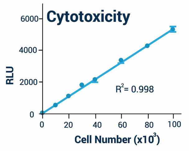
Plot the relative light units (RLU) against cell number to demonstrate linearity, and determine relative cell viability or cytotoxicity by comparing treated wells with untreated control wells.
| Component | Quantity | Storage |
| Assay Buffer | 10 mL | -20C |
| Substrate | 120 uL | -20C |
| ATP Enzyme | 120 uL | -20C |