The COX1 Antibody (CAB7531) is a high-quality antibody developed for reliable detection and analysis of target proteins. This antibody is produced in rabbits and is highly reactive with human samples, making it an ideal choice for Western blot applications. By binding specifically to the COX1 protein, this antibody enables accurate detection and analysis in a variety of cell types, making it a versatile tool for studies in immunology, inflammation, and cancer research.COX1 is a key enzyme in the inflammatory response and is known to play a role in various diseases, including cancer, cardiovascular disease, and inflammatory conditions.
This antibody is validated for use in WB, IHC-P, ELISA, IF applications and has demonstrated reactivity against Human, Mouse, Rat samples.
Product Name:
COX1 Antibody
SKU:
CAB7531
Size:
20μL, 100μL
Reactivity:
Human, Mouse, Rat
Conjugate:
Unconjugated
Immunogen:
Synthetic peptide. This information is considered to be commercially sensitive.
Enables cytochrome-c oxidase activity. Predicted to be involved in electron transport coupled proton transport; mitochondrial electron transport, cytochrome c to oxygen; and response to oxidative stress. Located in mitochondrial inner membrane. Part of mitochondrial respiratory chain complex IV. Is expressed in several structures, including brown fat; heart; liver; metanephros; and skeletal muscle. Orthologous to human MT-CO1 (mitochondrially encoded cytochrome c oxidase I).
Purification Method
Affinity purification
Gene ID
17708
RRID
AB_2768058
Buffer Information
Store at -20℃. Avoid freeze / thaw cycles. Buffer: PBS containing 50% glycerol, preserved with proclin300 or sodium azide, pH 7.3.
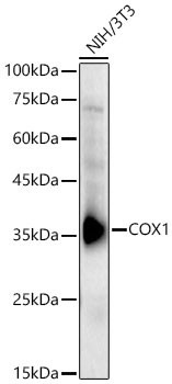
Western blot analysis of lysates from NIH/3T3 cells using COX1 Rabbit pAb (CAB7531) at 1:1800 dilution. Secondary antibody: HRP-conjugated Goat anti-Rabbit IgG (H+L) (CABS014) at 1:10000 dilution. Lysates/proteins: 25 μg per lane. Blocking buffer: 3% nonfat dry milk in TBST. Detection: ECL Basic Kit (AbGn00020). Exposure time:30s.
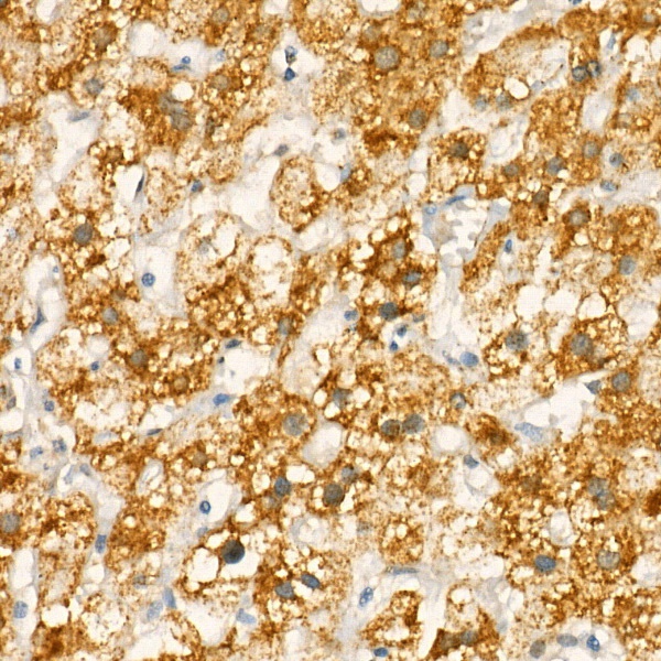
Immunohistochemistry analysis of paraffin-embedded Human liver using COX1 Rabbit pAb (CAB7531) at dilution of 1:20 (40x lens). High pressure antigen retrieval performed with 0.01M Citrate buffer (pH 6.0) prior to IHC staining.