The Phospho-(Ser/Thr) ATM/ATR Substrate Antibody (CABP0933) is a high-quality antibody developed for reliable detection and analysis of target proteins. This antibody, raised in rabbits, is highly sensitive and specific for detecting phospho-serine/threonine residues on ATM/ATR substrates in human samples. It has been validated for use in Western blot applications, allowing for the reliable detection and analysis of phospho-ser/thr ATM/ATR substrates in a variety of cell types.ATM (ataxia-telangiectasia mutated) and ATR (ATM and Rad3-related) are key kinases involved in DNA damage response pathways, regulating cell cycle checkpoints and DNA repair processes.
This antibody is validated for use in WB, ELISA applications and has demonstrated reactivity against Human, Mouse, Rat samples.
Product Name:
Phospho-(Ser/Thr) ATM/ATR Substrate Antibody
SKU:
CABP0933
Size:
20μL, 100μL
Reactivity:
Human, Mouse, Rat
Immunogen:
Synthetic peptide. This information is considered to be commercially sensitive.
Tested Applications:
WBELISA
Recommended Dilution:
WB
1:500 - 1:2000
ELISA
Recommended starting concentration is 1 μg/mL. Please optimize the concentration based on your specific assay requirements.
Positive Sample:
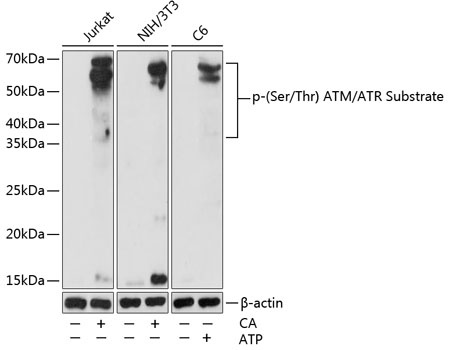
Jurkat treated with Calyculin A, NIH/3T3 treated with Calyculin A, C6 treated with ATP
Observed MW:
38-68kDa
The functionally related ATM (ataxia telangiectasia-mutated) and ATR (ATM-Rad3-related) protein kinases are critical regulators of DNA damage responses in mammalian cells. ATM and ATR share highly overlapping substrate specificities and show a strong preference for the phosphorylation of Serine (S) or Threonine (T) residues followed by Gln. It also called SQ or TQ consensus sites.
Purification Method
Affinity purification
RRID
AB_2863845
Buffer Information
Store at -20℃. Avoid freeze / thaw cycles. Buffer: PBS with 0.01% thimerosal,50% glycerol,pH7.3.
Western blot analysis of various lysates using Phospho-(Ser/Thr) ATM/ATR Substrate pAb (CABP0933) at 1:1000 dilution. Jurkat and NIH/3T3 cells were treated with Calyculin A (100 nM) at 37℃ for 30 minutes after serum-starvation overnight. C6 cells were treated with ATP(5 mM) at 30℃ for 1 hour. Secondary antibody: HRP-conjugated Goat anti-Rabbit IgG (H+L) (CABS014) at 1:10000 dilution. Lysates/proteins: 25μg per lane. Blocking buffer: 3% nonfat dry milk in TBST. Detection: ECL Basic Kit (AbGn00020). Exposure time: 60s.