The WNT9A Antibody (CAB7939) is a high-quality antibody developed for reliable detection and analysis of target proteins. This antibody, generated in rabbits, exhibits high specificity and sensitivity for human samples, making it an ideal choice for Western blot applications.By targeting the WNT-9A protein, this antibody allows for the detection and analysis of WNT-9A expression in various cell types, providing insights into its functions and interactions.
This antibody is validated for use in WB, ELISA applications and has demonstrated reactivity against Human, Mouse, Rat samples.
Product Name:
WNT9A Antibody
SKU:
CAB7939
Size:
20μL, 100μL
Reactivity:
Human, Mouse, Rat
Conjugate:
Unconjugated
Immunogen:
Recombinant protein (or fragment).This information is considered to be commercially sensitive.
The WNT gene family consists of structurally related genes that encode secreted signaling proteins. These proteins have been implicated in oncogenesis and in several developmental processes, including regulation of cell fate and patterning during embryogenesis. This gene is a member of the WNT gene family. It is expressed in gastric cancer cell lines. The protein encoded by this gene shows 75% amino acid identity to chicken Wnt14, which has been shown to play a central role in initiating synovial joint formation in the chick limb. This gene is clustered with another family member, WNT3A, in the chromosome 1q42 region.
Purification Method
Affinity purification
Gene ID
7483
RRID
AB_2772907
Buffer Information
Store at -20℃. Avoid freeze / thaw cycles. Buffer: PBS containing 50% glycerol, preserved with proclin300 or sodium azide, pH 7.3.
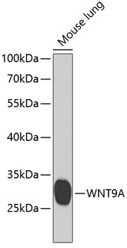
Western blot analysis of lysates from mouse lung, using WNT9A Rabbit pAb (CAB7939) at 1:1000 dilution. Secondary antibody: HRP-conjugated Goat anti-Rabbit IgG (H+L) (CABS014) at 1:10000 dilution. Lysates/proteins: 25μg per lane. Blocking buffer: 3% nonfat dry milk in TBST. Detection: ECL Basic Kit (AbGn00020). Exposure time: 90s.