The TCF4/TCF7L2 Monoclonal Antibody (CAB19548) is a high-quality antibody developed for reliable detection and analysis of target proteins. This antibody, generated in rabbits, is highly specific and reactive with human samples, making it ideal for use in Western blot applications.TCF7L2 is a key player in the Wnt signaling pathway, which regulates cell proliferation, differentiation, and survival. Dysregulation of TCF7L2 has been implicated in various diseases, including cancer and diabetes.
This antibody is validated for use in WB, IHC-P, ELISA applications and has demonstrated reactivity against Human samples.
Product Name:
TCF4/TCF7L2 Monoclonal Antibody
SKU:
CAB19548
Size:
20μL, 100μL
Reactivity:
Human
Clone Number:
ARC0027
Conjugate:
Unconjugated
Immunogen:
Synthetic peptide. This information is considered to be commercially sensitive.
Recommended starting concentration is 1 μg/mL. Please optimize the concentration based on your specific assay requirements.
Synonyms:
TCF4, TCF-4, TCF4/TCF7L2
Positive Sample:
SW620, HeLa, U-251MG, HepG2
Cellular Localization:
Nucleoplasm, Nucleus, Pml Body.
Calculated MW:
68kDa
Observed MW:
65kDa
This gene encodes a high mobility group (HMG) box-containing transcription factor that plays a key role in the Wnt signaling pathway. The protein has been implicated in blood glucose homeostasis. Genetic variants of this gene are associated with increased risk of type 2 diabetes. Several transcript variants encoding multiple different isoforms have been found for this gene.
Purification Method
Affinity purification
Gene ID
6934
RRID
AB_2862661
Buffer Information
Store at -20℃. Avoid freeze / thaw cycles. Buffer: PBS containing 50% glycerol and 0.05% BSA, preserved with proclin300 or sodium azide, pH 7.3.
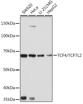
Western blot analysis of various lysates using TCF4/TCF7L2 Rabbit mAb (CAB19548) at 1:1000 dilution. Secondary antibody: HRP-conjugated Goat anti-Rabbit IgG (H+L) (CABS014) at 1:10000 dilution. Lysates/proteins: 25μg per lane. Blocking buffer: 3% nonfat dry milk in TBST. Detection: ECL Basic Kit (AbGn00020). Exposure time: 1s.
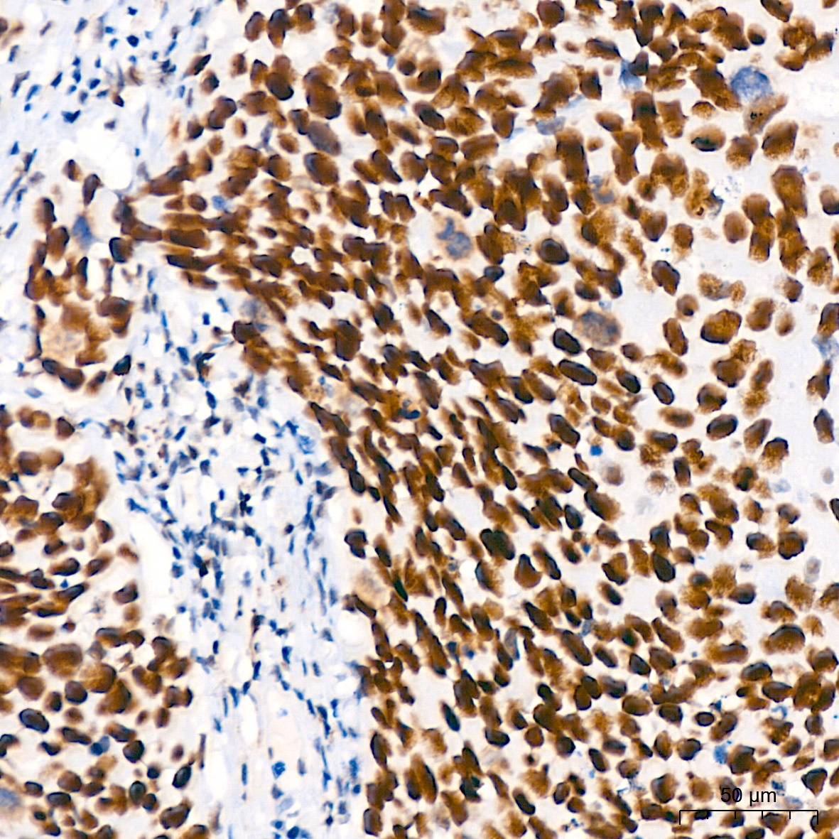
Immunohistochemistry analysis of paraffin-embedded Human cervix cancer tissue using TCF4/TCF7L2 Rabbit mAb (CAB19548) at a dilution of 1:200 (40x lens). High pressure antigen retrieval performed with 0.01M Tris-EDTA Buffer (pH 9.0) prior to IHC staining.
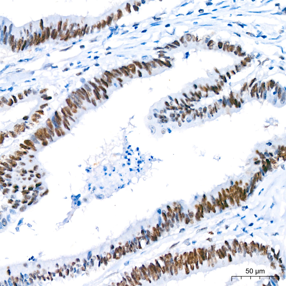
Immunohistochemistry analysis of paraffin-embedded Human colon carcinoma tissue using TCF4/TCF7L2 Rabbit mAb (CAB19548) at a dilution of 1:200 (40x lens). High pressure antigen retrieval performed with 0.01M Tris-EDTA Buffer (pH 9.0) prior to IHC staining.
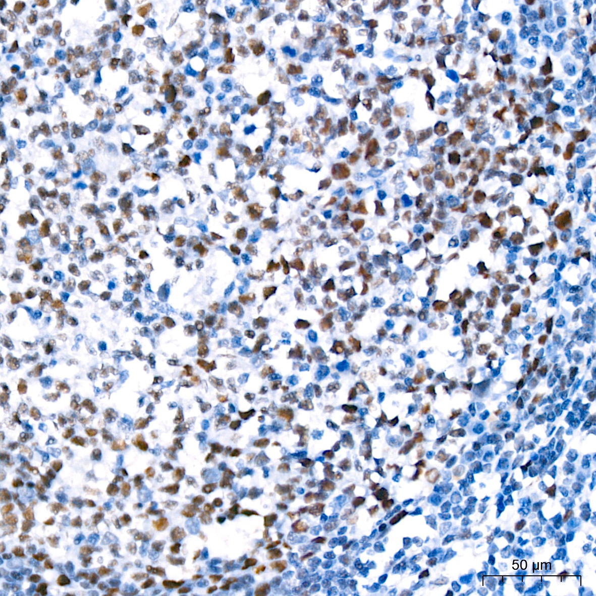
Immunohistochemistry analysis of paraffin-embedded Human tonsil tissue using TCF4/TCF7L2 Rabbit mAb (CAB19548) at a dilution of 1:200 (40x lens). High pressure antigen retrieval performed with 0.01M Citrate buffer (pH 6.0) prior to IHC staining.


 at 1:10000 dilution. Lysates/proteins: 25ug per lane. Blocking buffer: 3% nonfat dry milk in TBST. Detection: ECL Basic Kit. Exposure time: 180s.")

")


![Anti-TCF4 [R09-1D1] Monoclonal Antibody (AGMB00213)](https://cdn11.bigcommerce.com/s-h68l9z2lnx/images/stencil/590x590/products/271502/692470/anti-tcf4-r09-1d1-monoclonal-antibody-agmb00213__42537.1774506220.jpg?c=2 "Anti-TCF4 [R09-1D1] Monoclonal Antibody (AGMB00213)")

