Description
ADP Assay Kit (Luminescent) (BA0072) (BA0072)
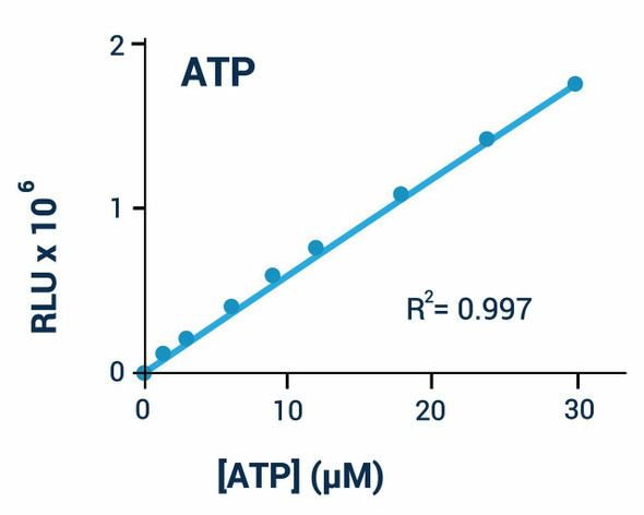
The ADP Assay Kit (Luminescent) (SKU: BA0072) provides a rapid bioluminescent method for measuring ADP levels. The assay involves two steps: in the first, a working reagent lyses cells to release ATP and ADP, and in the presence of luciferase the ATP reacts with the substrate D-luciferin to produce light whose intensity is a direct measure of ATP. In the second step, the ADP is converted to ATP through an enzyme reaction, and the newly formed ATP produces light as before; the second measurement represents the total ADP plus ATP. This non-radioactive, homogeneous cell-based assay is performed in microplates and is compatible with all culture media and liquid handling systems for high-throughput screening in 96-well and 384-well plates.
| Product Name: | ADP Assay Kit (Luminescent) (BA0072) |
| SKU: | BA0072 |
| Detection Method: | Luminescent (bioluminescent) |
| Detection Range: | As low as 0.02 uM; up to 30 uM ADP |
| Sample Type: | Cells, tissue and other biological samples |
| Species Reactivity: | All |
| Assay Time: | Approximately 12 minutes (10 minute ATP reaction plus 2 minute ADP reaction) |
| Kit Size: | 100 Assays |
| Equipment Required: | Microplate reader |
| Storage: | -20C |
| Shelf Life: | 12 months after receipt |
| Shipping: | Gel Pack |
Rapid bioluminescent determination of ADP. Luciferase converts ATP and D-luciferin to light; ADP is then converted to ATP and measured a second time, with the difference reflecting the ADP concentration.
- Safe, non-radioactive assay
- Sensitive: as low as 0.02 uM ADP can be quantified
- Homogeneous mix-incubate-measure format with no wash or transfer steps
- Robust and amenable to high-throughput screening, with Z' factors of 0.5 and above routinely observed
- ADP determination in cells and other biological samples
Note: The below protocol is a sample protocol. Protocols are specific to each batch/lot. For the correct instructions please follow the protocol included in your kit.
| Step | Procedure |
| 1 | Standard curve. Prepare 500 uL 30 uM ADP premix by mixing 5 uL 3 mM Standard with 495 uL distilled water (or culture media for cell samples) and prepare the dilution series shown in the table. Transfer 10 uL standards into wells of a white opaque 96-well plate. Use 10 uL sample per well; for tissue, homogenise 20 mg in 200 uL cold PBS, spin at 12,000 g for 5 minutes and use supernatant; for suspension cells transfer 10 uL of 1,000-10,000 cells; for adherent cells culture 1,000-10,000 cells and remove medium before adding ATP Reagent. |
| 2 | ATP reaction. Bring Assay Buffer, Substrate and Cosubstrate to room temperature and thaw enzyme on ice. For each well mix 95 uL Assay Buffer with 1 uL Substrate, 1 uL Cosubstrate and 1 uL ATP Enzyme. Add 90 uL ATP Reagent to each well, mix by tapping and after 10 minutes read luminescence (RLU A). |
| 3 | ADP assay. Prepare ADP Reagent by mixing, for each well, 5 uL distilled water with 1 uL ADP Enzyme. Immediately after reading RLU A, add 5 uL ADP Reagent to each well, mix, incubate 2 minutes at room temperature and read luminescence (RLU B). |
Subtract RLU A from RLU B for standards and samples. Plot the change in RLU against ADP concentration for the standards and determine the slope. [ADP]sample = ((RLU B)sample - (RLU A)sample) / slope uM.
| Component | Quantity | Storage |
| Assay Buffer | 10 mL | -20C |
| Cosubstrate | 120 uL | -20C |
| ADP Enzyme | 120 uL | -20C |
| Substrate | 120 uL | -20C |
| ATP Enzyme | 120 uL | -20C |
| Standard (3 mM ADP) | 100 uL | -20C |