The EHMT2 Monoclonal Antibody (CAB19288) is a high-quality antibody developed for reliable detection and analysis of target proteins. This antibody is raised in rabbits and has been validated for use in various applications, including Western blot and immunohistochemistry.EHMT2, also known as G9a, plays a critical role in gene expression regulation through histone methylation, impacting various cellular processes such as differentiation, development, and disease progression. The Anti-EHMT2 Antibody specifically binds to EHMT2, enabling precise detection and analysis in different cell types, making it a valuable tool for researchers in the fields of epigenetics, cancer biology, and developmental biology.
This antibody is validated for use in WB, IHC-P, ELISA applications and has demonstrated reactivity against Human, Mouse, Rat samples.
Product Name:
EHMT2 Monoclonal Antibody
SKU:
CAB19288
Size:
20μL, 100μL
Reactivity:
Human, Mouse, Rat
Clone Number:
ARC2470
Conjugate:
Unconjugated
Immunogen:
Recombinant protein (or fragment).This information is considered to be commercially sensitive.
Recommended starting concentration is 1 μg/mL. Please optimize the concentration based on your specific assay requirements.
Synonyms:
G9A, BAT8, GAT8, NG36, KMT1C, C6orf30, EHMT2
Positive Sample:
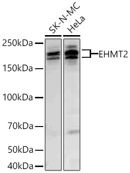
SK-N-MC, HeLa
Cellular Localization:
Chromosome, Nucleus.
Calculated MW:
132kDa
Observed MW:
160kDa/180kDa
This gene encodes a methyltransferase that methylates lysine residues of histone H3. Methylation of H3 at lysine 9 by this protein results in recruitment of additional epigenetic regulators and repression of transcription. This gene was initially thought to be two different genes, NG36 and G9a, adjacent to each other in the HLA locus. Alternative splicing results in multiple transcript variants.
Purification Method
Affinity purification
Gene ID
10919
Buffer Information
Store at -20℃. Avoid freeze / thaw cycles. Buffer: PBS containing 50% glycerol and 0.05% BSA, preserved with proclin300 or sodium azide, pH 7.3.
Western blot analysis of various lysates using EHMT2 Rabbit mAb (CAB19288) at 1:5000 dilution incubated overnight at 4℃. Secondary antibody: HRP-conjugated Goat anti-Rabbit IgG (H+L) (CABS014) at 1:10000 dilution. Lysates/proteins: 25 μg per lane. Blocking buffer: 3% nonfat dry milk in TBST. Detection: ECL Basic Kit (AbGn00020). Exposure time: 90 s.
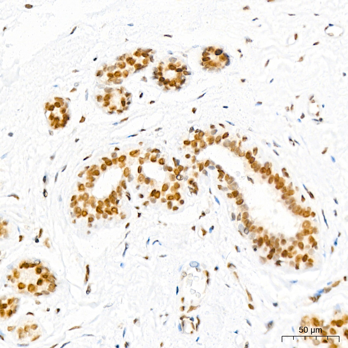
Immunohistochemistry analysis of paraffin-embedded Human breast tissue using EHMT2 Rabbit mAb (CAB19288) at a dilution of 1:200 (40x lens). High pressure antigen retrieval performed with 0.01M Citrate buffer (pH 6.0) prior to IHC staining.
Immunohistochemistry analysis of paraffin-embedded Human thyroid tissue using EHMT2 Rabbit mAb (CAB19288) at a dilution of 1:200 (40x lens). High pressure antigen retrieval performed with 0.01M Citrate buffer (pH 6.0) prior to IHC staining.
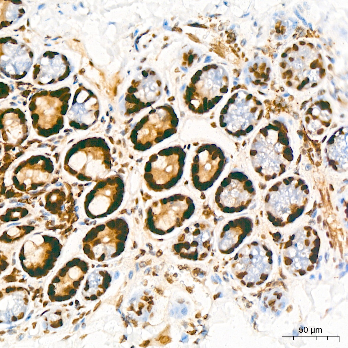
Immunohistochemistry analysis of paraffin-embedded Mouse colon tissue using EHMT2 Rabbit mAb (CAB19288) at a dilution of 1:200 (40x lens). High pressure antigen retrieval performed with 0.01M Citrate buffer (pH 6.0) prior to IHC staining.
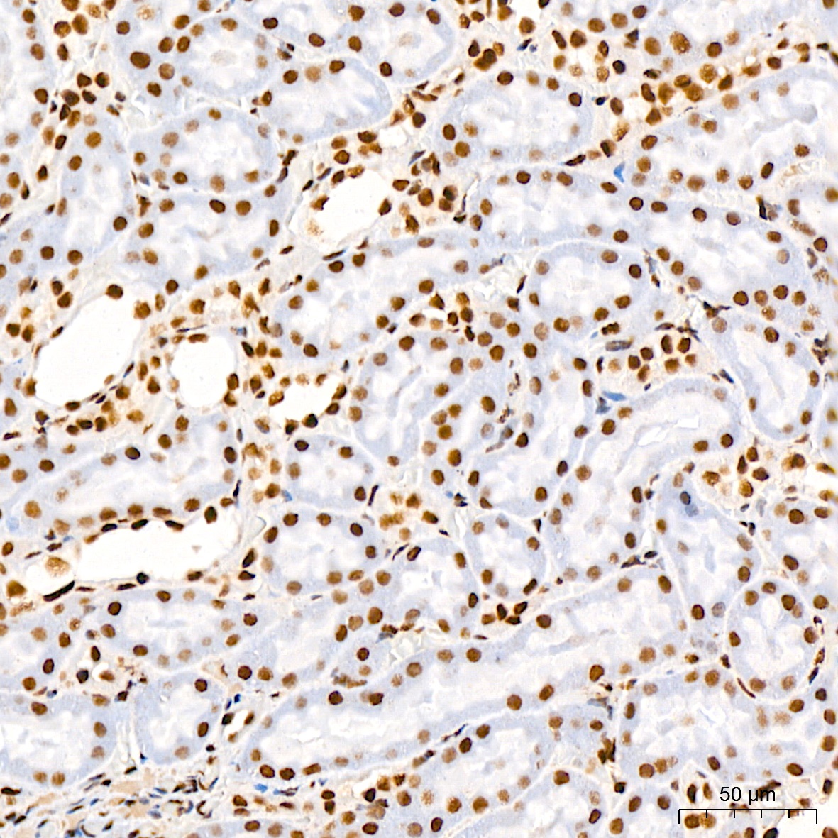
Immunohistochemistry analysis of paraffin-embedded Rat kidney tissue using EHMT2 Rabbit mAb (CAB19288) at a dilution of 1:200 (40x lens). High pressure antigen retrieval performed with 0.01M Citrate buffer (pH 6.0) prior to IHC staining.
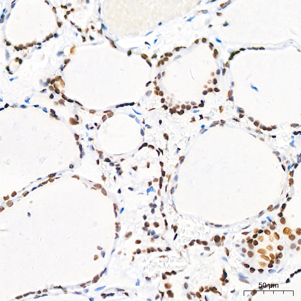
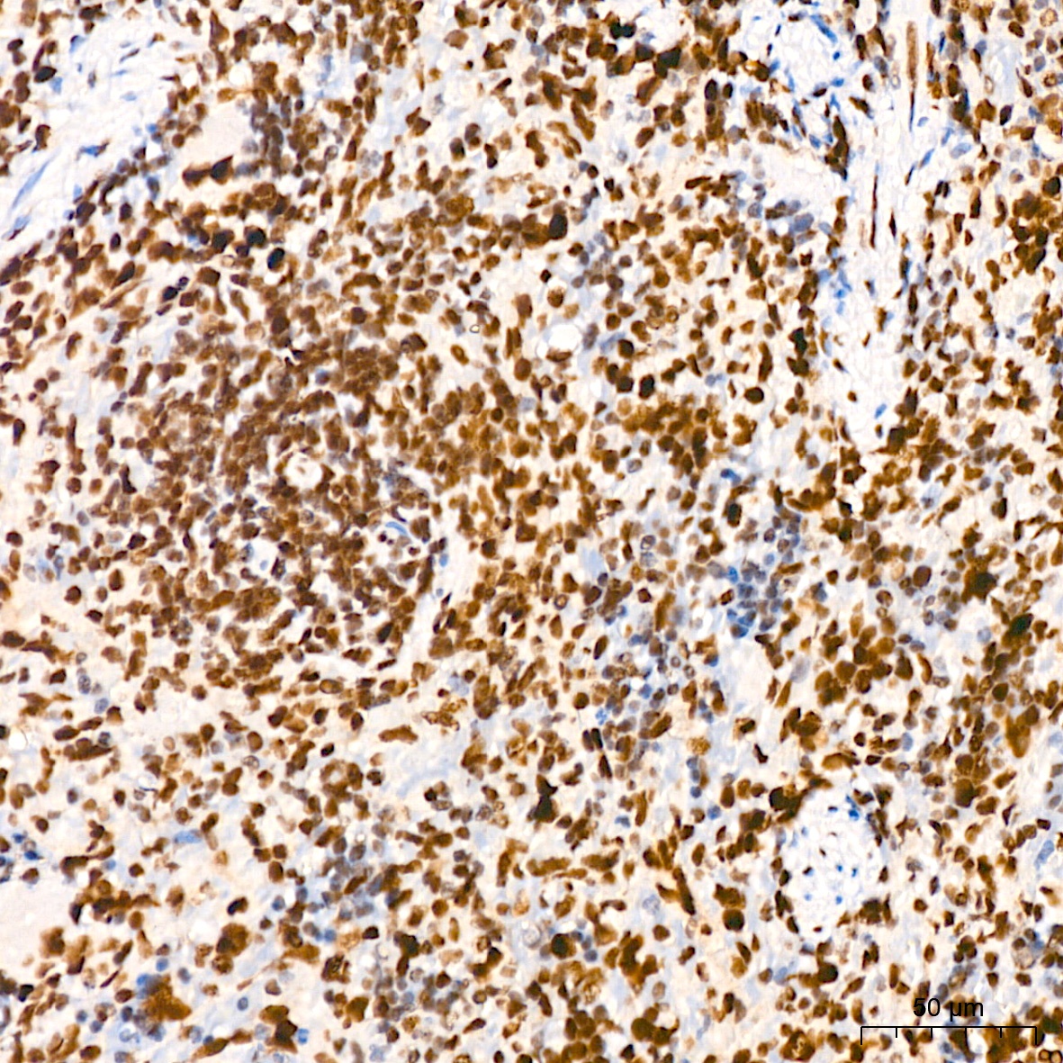
Immunohistochemistry analysis of paraffin-embedded Rat spleen tissue using EHMT2 Rabbit mAb (CAB19288) at a dilution of 1:200 (40x lens). High pressure antigen retrieval performed with 0.01M Citrate buffer (pH 6.0) prior to IHC staining.


![Anti-EHMT2 [R03-5U-1] Monoclonal Antibody (AGMB03865)](https://cdn11.bigcommerce.com/s-h68l9z2lnx/images/stencil/590x590/products/275154/680847/anti-ehmt2-r03-5u-1-monoclonal-antibody-agmb03865__39438.1773043352.jpg?c=2 "Anti-EHMT2 [R03-5U-1] Monoclonal Antibody (AGMB03865)")
![Anti-EHMT2 [R05-8H2] Monoclonal Antibody (AGMB00388)](https://cdn11.bigcommerce.com/s-h68l9z2lnx/images/stencil/590x590/products/271677/693292/anti-ehmt2-r05-8h2-monoclonal-antibody-agmb00388__00045.1774508848.jpg?c=2 "Anti-EHMT2 [R05-8H2] Monoclonal Antibody (AGMB00388)")


