The APRT Antibody (CAB5456) is a high-quality antibody developed for reliable detection and analysis of target proteins. This antibody, raised in rabbits, shows high reactivity with human samples and is validated for use in Western blot applications. By specifically binding to the APRT protein, this antibody allows for precise detection and analysis in a variety of cell types, making it a valuable asset for studies in biochemistry and molecular biology.APRT, or adenine phosphoribosyltransferase, plays a crucial role in the recycling of purine bases in the body, contributing to nucleotide synthesis and maintaining cellular energy balance.
This antibody is validated for use in WB, ELISA applications and has demonstrated reactivity against Human, Mouse samples.
Product Name:
APRT Antibody
SKU:
CAB5456
Size:
20μL, 100μL
Reactivity:
Human, Mouse
Conjugate:
Unconjugated
Immunogen:
Recombinant protein (or fragment).This information is considered to be commercially sensitive.
Recommended starting concentration is 1 μg/mL. Please optimize the concentration based on your specific assay requirements.
Synonyms:
AMP, APRTD, APRT
Positive Sample:
293T
Cellular Localization:
Cytoplasm.
Calculated MW:
20kDa
Observed MW:
22kDa
Adenine phosphoribosyltransferase belongs to the purine/pyrimidine phosphoribosyltransferase family. A conserved feature of this gene is the distribution of CpG dinucleotides. This enzyme catalyzes the formation of AMP and inorganic pyrophosphate from adenine and 5-phosphoribosyl-1-pyrophosphate (PRPP). It also produces adenine as a by-product of the polyamine biosynthesis pathway. A homozygous deficiency in this enzyme causes 2,8-dihydroxyadenine urolithiasis. Two transcript variants encoding different isoforms have been found for this gene.
Purification Method
Affinity purification
Gene ID
353
RRID
AB_2766257
Buffer Information
Store at -20℃. Avoid freeze / thaw cycles. Buffer: PBS containing 50% glycerol, preserved with proclin300 or sodium azide, pH 7.3.
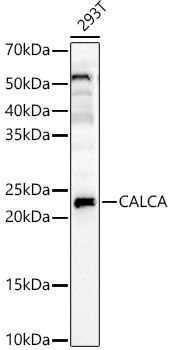
Western blot analysis of lysates from 293T cells, using CALCA Rabbit pAb (CAB5456) at 1:2500 dilution. Secondary antibody: HRP-conjugated Goat anti-Rabbit IgG (H+L) (CABS014) at 1:10000 dilution. Lysates/proteins: 25μg per lane. Blocking buffer: 3% nonfat dry milk in TBST. Detection: ECL Enhanced Kit (AbGn00021). Exposure time: 30s.