
JAK-STAT Signalling and Cytokines: The What, How, and Why
JAK-STAT Signalling: Mechanism, Regulation and Inhibitors
JAK-STAT is the shortest route from a cytokine outside the cell to a change in gene transcription — receptor engagement, kinase activation, transcription factor phosphorylation, nuclear entry. There are only four JAKs and seven STATs, yet they carry signalling for most interleukins, all interferons and several growth factors. This guide covers the mechanism, the negative feedback that limits it, and the inhibitors built to block it.
Browse JAK-STAT antibodies →Key takeaways
- There are four JAKs: JAK1, JAK2, JAK3 and TYK2. TYK2 is not called JAK4 — no such protein exists.
- Cytokine receptors have no intrinsic kinase activity. Neither type I nor type II does. That is precisely why they depend on non-covalently associated JAKs, and it is the defining feature of the receptor class.
- Unphosphorylated STATs are predominantly cytoplasmic monomers. Phosphorylation drives dimerisation through reciprocal SH2-phosphotyrosine binding, and that dimerisation is the activation step.
- The pathway is unusually short — receptor to nucleus with no second messenger and no kinase cascade — which is why it is fast and why it needs strong negative regulation.
- SOCS proteins provide that brake: they are themselves STAT target genes, forming a classic negative feedback loop.
- JAK2 V617F is the defining mutation of the myeloproliferative neoplasms and the reason ruxolitinib exists.
- JAK inhibitors are effective in rheumatoid arthritis, psoriasis, IBD and alopecia areata, and carry class boxed warnings for serious infection, thrombosis, major cardiovascular events and malignancy.
JAK-STAT antibodies
The kinases, the principal STATs, the phospho-site that reports activation, and SOCS3 for the feedback arm.
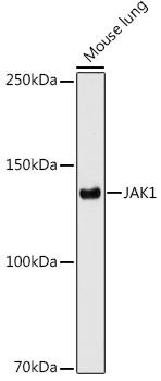
JAK1 Rabbit Monoclonal Antibody
Pairs with almost every other JAK; used by the gp130, common gamma chain and interferon receptor families.
View product →![Anti-JAK2 [R03-1F2] Monoclonal Antibody](https://cdn11.bigcommerce.com/s-h68l9z2lnx/products/273193/images/678194/anti-jak2-r03-1f2-monoclonal-antibody-agmb01904__04841.1773034958.386.513.jpg?c=2)
Anti-JAK2 [R03-1F2] Monoclonal Antibody
Carries the V617F mutation of the myeloproliferative neoplasms and serves the erythropoietin and growth hormone receptors.
View product →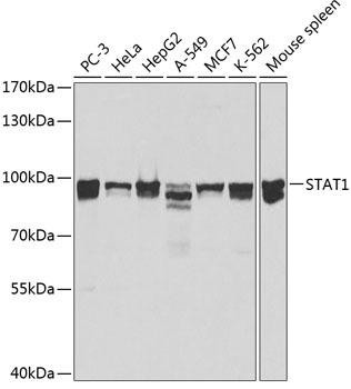
STAT1 Rabbit Polyclonal Antibody
The interferon-responsive STAT; central to antiviral and antimycobacterial immunity.
View product →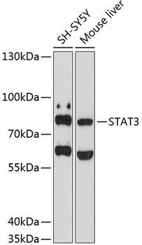
STAT3 Rabbit Polyclonal Antibody
The IL-6 family STAT, constitutively active in many tumours and mutated in hyper-IgE syndrome.
View product →
Anti-Phospho-STAT3 (Tyr705) Monoclonal Antibody
Tyr705 phosphorylation is the activation event — the direct readout of pathway engagement.
View product →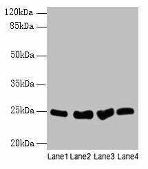
SOCS3 Antibody
The induced feedback inhibitor; measuring it reports whether the brake has engaged.
View product →JAK proteins
JAK stands for Janus kinase, named after the two-faced Roman god because each enzyme carries two kinase-like domains. All four are non-receptor tyrosine kinases that sit constitutively bound to the cytoplasmic tails of cytokine receptors.
| Kinase | Principal receptor partners | Consequence of loss |
|---|---|---|
| JAK1 | Common gamma chain, gp130 and interferon receptor families | Broad; embryonically or perinatally lethal in mice |
| JAK2 | Erythropoietin, thrombopoietin, growth hormone, IL-3 family | Failure of erythropoiesis |
| JAK3 | Exclusively the common gamma chain receptors | Severe combined immunodeficiency — its restricted expression makes it an attractive selective target |
| TYK2 | Type I interferon, IL-12 and IL-23 receptors | Susceptibility to mycobacteria and viruses; loss-of-function variants protect against autoimmunity |
To be explicit on a common error: the fourth kinase is TYK2, and it is not designated JAK4. No protein of that name exists.
Each JAK has a FERM domain and an SH2-like domain that mediate receptor binding, followed by a pseudokinase domain and a catalytically active kinase domain. The pseudokinase domain is not merely vestigial — it autoinhibits the active domain, and the V617F mutation of JAK2 falls within it, relieving that inhibition.
STAT proteins
STAT stands for signal transducer and activator of transcription — a name that captures the unusual point that one protein performs both roles. Seven exist in mammals, from six genes: STAT1, STAT2, STAT3, STAT4, STAT5A, STAT5B and STAT6.
Each has an N-terminal domain, coiled-coil domain, DNA-binding domain, linker, SH2 domain and a C-terminal transactivation domain, with a single critical tyrosine between the SH2 and transactivation domains.
Monomer, not pre-formed active dimer
Unphosphorylated STATs are predominantly cytoplasmic monomers. On phosphorylation of that critical tyrosine, the SH2 domain of one STAT binds the phosphotyrosine of another, and the reciprocal interaction locks them into a parallel dimer that exposes a nuclear localisation signal.
Dimerisation is therefore the activation step, not a pre-existing state. Some unphosphorylated STAT does exist as antiparallel dimers with distinct functions, but describing STATs as sitting in the cytoplasm as inactive dimers omits the mechanism that actually switches the pathway on.
The pathway step by step
- Ligand binding. A cytokine binds its receptor, bringing two or more receptor chains — and their associated JAKs — into proximity.
- JAK activation. The apposed JAKs trans-phosphorylate one another on activation-loop tyrosines, relieving pseudokinase autoinhibition.
- Receptor phosphorylation. Active JAKs phosphorylate tyrosines on the receptor cytoplasmic tail, creating SH2 docking sites.
- STAT recruitment. STATs dock at those receptor phosphotyrosines through their own SH2 domains. Which STAT is recruited depends on the sequence around each site, and this is where much of the pathway’s specificity resides.
- STAT phosphorylation and dimerisation. JAKs phosphorylate the critical STAT tyrosine; two phosphorylated STATs form a reciprocal SH2-phosphotyrosine dimer.
- Nuclear translocation and transcription. The dimer enters the nucleus and binds GAS or ISRE elements, activating or repressing target genes.
What is notable is the absence of anything in between. No second messenger, no kinase cascade — the receptor and the transcription factor are separated by a single phosphorylation event. That directness makes the pathway fast, and makes tight negative regulation essential.
Cytokine receptors
An important correction to a common misstatement: cytokine receptors possess no intrinsic kinase activity of their own. This applies to both type I and type II. It is the defining property of the class and the entire reason JAKs are required — a receptor tyrosine kinase such as EGFR phosphorylates its own tail, whereas a cytokine receptor cannot and must borrow a kinase.
| Class | Structural feature | Examples |
|---|---|---|
| Type I (haematopoietin) | Conserved cysteines and a WSXWS motif in the extracellular domain | IL-2 to IL-7, IL-12, IL-15, IL-21, GM-CSF, erythropoietin, growth hormone |
| Type II | Related fold but lacking the WSXWS motif | All interferons, IL-10 family |
Both classes signal identically in mechanism — associated JAKs, receptor phosphorylation, STAT recruitment. The type I/type II division is structural, not functional.
Many receptors also share subunits: the common gamma chain across IL-2, 4, 7, 9, 15 and 21, and gp130 across the IL-6 family. Shared subunits explain both the redundancy between cytokines and the breadth of effect when one is blocked.
Which cytokine uses which JAK and STAT
| Cytokine | JAKs | Principal STATs | Main outcome |
|---|---|---|---|
| IL-6 family | JAK1, JAK2, TYK2 | STAT3 | Acute phase response; Th17 differentiation |
| Type I interferons | JAK1, TYK2 | STAT1, STAT2 with IRF9 | Antiviral gene programme |
| Interferon gamma | JAK1, JAK2 | STAT1 | Macrophage activation; MHC upregulation |
| IL-2, IL-15 | JAK1, JAK3 | STAT5 | T and NK cell proliferation |
| IL-4, IL-13 | JAK1, JAK3, TYK2 | STAT6 | Th2 differentiation; IgE class switching |
| IL-12 | JAK2, TYK2 | STAT4 | Th1 differentiation |
| Erythropoietin | JAK2 | STAT5 | Erythropoiesis |
These pairings explain inhibitor selectivity. A JAK3-sparing inhibitor leaves common gamma chain signalling largely intact; a TYK2-selective agent targets IL-12 and IL-23 without touching erythropoietin, which depends on JAK2.
Negative regulation
Because the pathway is so direct, it requires strong brakes. Three mechanisms operate, and the original article omitted all of them.
- SOCS proteins. The suppressor of cytokine signalling family are themselves STAT target genes — so activating the pathway induces its own inhibitor. SOCS bind phosphotyrosines on the receptor, block STAT docking, inhibit JAK catalysis directly, and recruit an E3 ubiquitin ligase to degrade the complex. A textbook negative feedback loop.
- Protein tyrosine phosphatases — SHP1, SHP2, CD45 and PTP1B — remove the activating phosphates from JAKs, receptors and STATs.
- PIAS proteins act in the nucleus, blocking DNA binding by activated STAT dimers.
Loss of these brakes matters: SOCS silencing is a recognised route to constitutive STAT3 activation in tumours, achieving the same end as an activating mutation.
Disease associations
| Condition | Molecular basis |
|---|---|
| Myeloproliferative neoplasms | JAK2 V617F in the pseudokinase domain causes ligand-independent activation — present in most polycythaemia vera and around half of essential thrombocythaemia and myelofibrosis |
| Severe combined immunodeficiency | Loss-of-function JAK3 or common gamma chain mutations abolish IL-2, 7 and 15 signalling |
| Autosomal dominant hyper-IgE (Job) syndrome | STAT3 loss-of-function — not STAT1, which is a frequent misattribution |
| Chronic mucocutaneous candidiasis | STAT1 gain-of-function, impairing Th17 responses |
| Mycobacterial and viral susceptibility | STAT1 loss-of-function, blocking interferon responses |
| Solid tumours and lymphoma | Constitutive STAT3 or STAT5 activation supporting proliferation, survival and immune evasion |
The STAT1 versus STAT3 distinction in the immunodeficiencies is worth holding onto, since the two are readily confused and point to entirely different clinical pictures.
JAK inhibitors
JAK inhibitors are small molecules that occupy the ATP-binding site of the kinase domain, preventing JAKs from phosphorylating the receptor and STATs. Several are approved, differing in which JAKs they hit.
| Agent | Selectivity | Principal uses |
|---|---|---|
| Ruxolitinib | JAK1 / JAK2 | Myelofibrosis, polycythaemia vera, graft-versus-host disease |
| Tofacitinib | Pan-JAK, JAK1 / JAK3 preference | Rheumatoid arthritis, psoriatic arthritis, ulcerative colitis |
| Baricitinib | JAK1 / JAK2 | Rheumatoid arthritis, alopecia areata, severe COVID-19 |
| Upadacitinib | JAK1-selective | Rheumatoid arthritis, atopic dermatitis, inflammatory bowel disease |
| Deucravacitinib | TYK2-selective, allosteric | Plaque psoriasis — binds the pseudokinase domain rather than the ATP site |
Safety
As a class these agents carry boxed warnings for serious infection (including tuberculosis reactivation and herpes zoster), venous thromboembolism, major adverse cardiovascular events and malignancy. Because JAK2 also serves erythropoietin and thrombopoietin, cytopenias are a predictable on-target effect.
A correction on mechanism: JAK inhibition reduces signalling by cytokines that use this pathway — IL-6, the interferons, IL-2, IL-12 and IL-23. TNF-alpha is not a JAK-STAT cytokine; it signals through NF-kappa-B and MAPK, so it is a poor example of what JAK inhibitors block. IL-6 is the appropriate one.
Clinical decisions rest with the treating clinician; this is research context only.
Measuring pathway activity
- Phospho-STAT by western blot or flow cytometry is the standard readout. Phospho-STAT3 at Tyr705 and phospho-STAT1 at Tyr701 are the usual targets, always normalised to total STAT.
- Timing matters. STAT phosphorylation peaks within 15 to 30 minutes of stimulation and declines as SOCS is induced, so a late time point can look like no response at all.
- SOCS induction at the mRNA or protein level confirms the pathway ran to completion rather than merely being triggered.
- Phospho-flow resolves responses cell by cell, which matters in mixed populations where different cells express different receptor combinations.
- Target gene readouts — interferon-stimulated genes, acute phase proteins — report the transcriptional consequence rather than the signalling event.
Choosing antibodies
JAK1 and JAK2, STAT1 and STAT3, phospho-STAT3 (Tyr705) as the activation readout, and SOCS3 for the feedback arm.
Browse JAK-STAT antibodies →Frequently asked questions
How many JAK proteins are there, and is TYK2 the same as JAK4?
Four: JAK1, JAK2, JAK3 and TYK2. TYK2 is not called JAK4 — there is no protein of that name, despite the designation appearing in some secondary sources.
Do cytokine receptors have their own kinase activity?
No. Neither type I nor type II cytokine receptors have intrinsic kinase activity, which is exactly why they depend on non-covalently associated JAKs. This distinguishes them from receptor tyrosine kinases such as EGFR, which phosphorylate their own tails.
Are STATs dimers before they are activated?
Predominantly they are cytoplasmic monomers. Phosphorylation of a single conserved tyrosine drives dimerisation through reciprocal SH2-phosphotyrosine binding, and that dimerisation exposes the nuclear localisation signal — so it is the activation step, not a pre-existing state.
What switches the pathway off?
Chiefly SOCS proteins, which are themselves STAT target genes and so form a negative feedback loop. Phosphatases including SHP1, SHP2 and CD45 remove activating phosphates, and PIAS proteins block DNA binding in the nucleus.
What is JAK2 V617F?
A point mutation in the JAK2 pseudokinase domain that relieves autoinhibition and causes ligand-independent activation. It is present in most cases of polycythaemia vera and around half of essential thrombocythaemia and myelofibrosis, and it is the rationale for ruxolitinib.
Which STAT is mutated in hyper-IgE syndrome?
STAT3. Autosomal dominant hyper-IgE (Job) syndrome results from STAT3 loss-of-function. STAT1 defects cause different pictures — gain-of-function gives chronic mucocutaneous candidiasis, loss-of-function gives mycobacterial and viral susceptibility.
Do JAK inhibitors block TNF-alpha?
Not directly. TNF-alpha signals through NF-kappa-B and MAPK rather than JAK-STAT. JAK inhibitors block cytokines that use this pathway — IL-6, the interferons, IL-2, IL-12 and IL-23 — so IL-6 is the appropriate example.
Recent Posts
-
Suspected Plague Death at a Siberian Anti-Plague Institute: What Researchers Should Know
Infectious Disease · News AnalysisSuspected Plague Death at a Siberian Anti-Plague Institute: What …6th Oct 2026 -
LPS ELISA Troubleshooting: The Importance of Sample Nature
Quick answer A researcher measuring circulating lipopolysaccharide (LPS) in human serum …25th Sep 2026 -

Sekundärantikörper wählen: Die Regel, die im Labor wirklich zählt
Sekundärantikörper wählen: Die Regel, die im Labor wirklich zählt Wählen Sie einen Sekundärantikörp …24th Sep 2026