The CSK Antibody (CAB16824) is a high-quality antibody developed for reliable detection and analysis of target proteins. Raised in rabbits, this antibody is highly specific and reactive with human samples, making it an ideal choice for Western blot applications. By binding to CSK, this antibody enables the detection and analysis of the protein in different cell types, allowing for the investigation of its role in cellular signaling and disease processes.CSK is known to play a crucial role in the regulation of cell proliferation, differentiation, and apoptosis.
This antibody is validated for use in WB, IHC-P, IF/ICC, ELISA applications and has demonstrated reactivity against Human, Mouse, Rat samples.
Product Name:
CSK Antibody
SKU:
CAB16824
Size:
20μL, 100μL
Reactivity:
Human, Mouse, Rat
Immunogen:
Recombinant protein (or fragment).This information is considered to be commercially sensitive.
Recommended starting concentration is 1 μg/mL. Please optimize the concentration based on your specific assay requirements.
Synonyms:
CSK
Positive Sample:
U-87MG, Mouse spleen
Cellular Localization:
Cell Membrane, Cytoplasm.
Calculated MW:
51kDa
Observed MW:
50kDa
The protein encoded by this gene is involved in multiple pathways, including the regulation of Src family kinases. It plays an important role in T-cell activation through its association with the protein encoded by the protein tyrosine phosphatase, non-receptor type 22 (PTPN22) gene. This protein also phosphorylates C-terminal tyrosine residues on multiple substrates, including the protein encoded by the SRC proto-oncogene, non-receptor tyrosine kinase gene. Phosphorylation suppresses the kinase activity of the Src family tyrosine kinases. An intronic polymorphism (rs34933034) in this gene has been found to affect B-cell activation and is associated with systemic lupus erythematosus (SLE). Alternative splicing results in multiple transcript variants.
Purification Method
Affinity purification
Gene ID
1445
RRID
AB_2769059
Buffer Information
Store at -20℃. Avoid freeze / thaw cycles. Buffer: PBS containing 50% glycerol, preserved with proclin300 or sodium azide, pH 7.3.
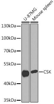
Western blot analysis of various lysates using CSK Rabbit pAb (CAB16824) at 1:1000 dilution. Secondary antibody: HRP-conjugated Goat anti-Rabbit IgG (H+L) (CABS014) at 1:10000 dilution. Lysates/proteins: 25μg per lane. Blocking buffer: 3% nonfat dry milk in TBST. Detection: ECL Basic Kit (AbGn00020). Exposure time: 90s.
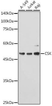
Western blot analysis of various lysates using CSK Rabbit pAb (CAB16824) at 1:1000 dilution. Secondary antibody: HRP-conjugated Goat anti-Rabbit IgG (H+L) (CABS014) at 1:10000 dilution. Lysates/proteins: 25μg per lane. Blocking buffer: 3% nonfat dry milk in TBST. Detection: ECL Basic Kit (AbGn00020). Exposure time: 60s.
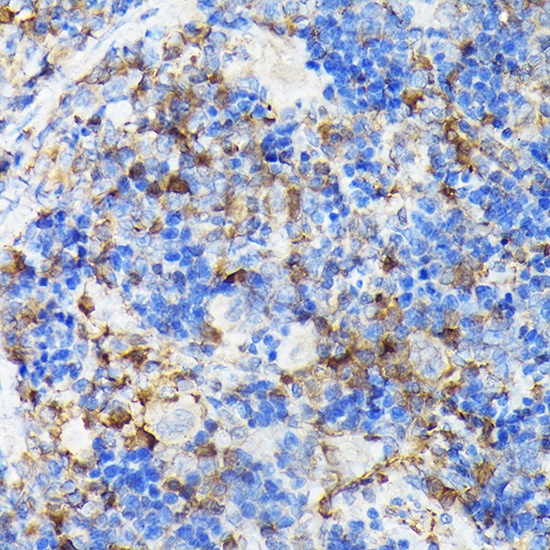
Immunohistochemistry analysis of paraffin-embedded Rat spleen using CSK Rabbit pAb (CAB16824) at dilution of 1:100 (40x lens). High pressure antigen retrieval performed with 0.01M Citrate buffer (pH 6.0) prior to IHC staining.
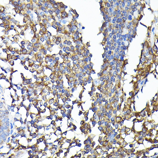
Immunohistochemistry analysis of paraffin-embedded Human colon carcinoma using CSK Rabbit pAb (CAB16824) at dilution of 1:100 (40x lens). High pressure antigen retrieval performed with 0.01M Citrate buffer (pH 6.0) prior to IHC staining.
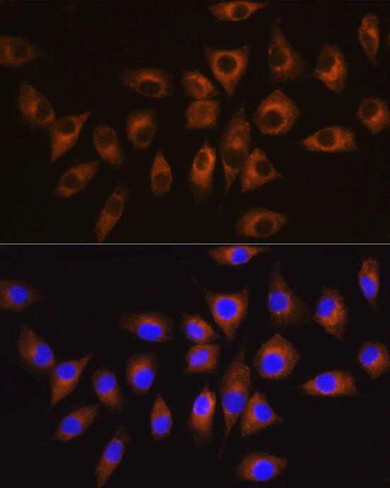
Immunofluorescence analysis of L929 cells using CSK Rabbit pAb (CAB16824) at dilution of 1:100 (40x lens). Secondary antibody: Cy3-conjugated Goat anti-Rabbit IgG (H+L) (CABS007) at 1:500 dilution. Blue: DAPI for nuclear staining.
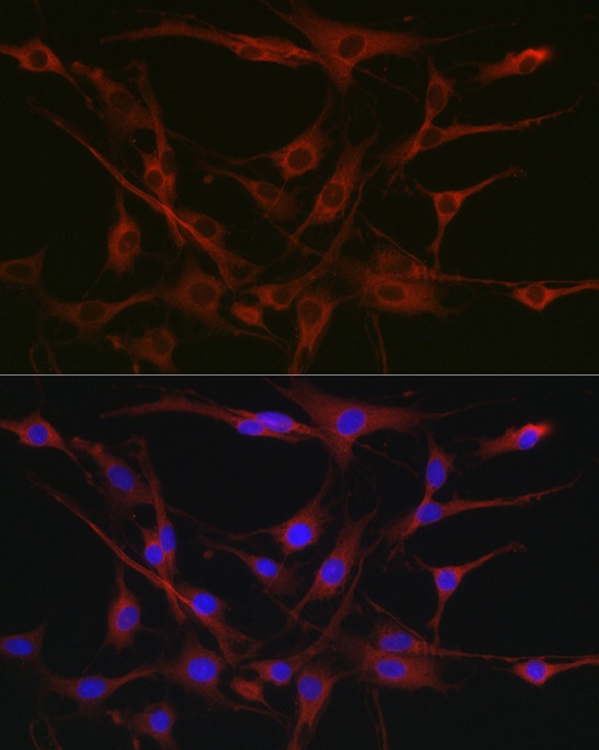
Immunofluorescence analysis of C6 cells using CSK Rabbit pAb (CAB16824) at dilution of 1:100 (40x lens). Secondary antibody: Cy3-conjugated Goat anti-Rabbit IgG (H+L) (CABS007) at 1:500 dilution. Blue: DAPI for nuclear staining.
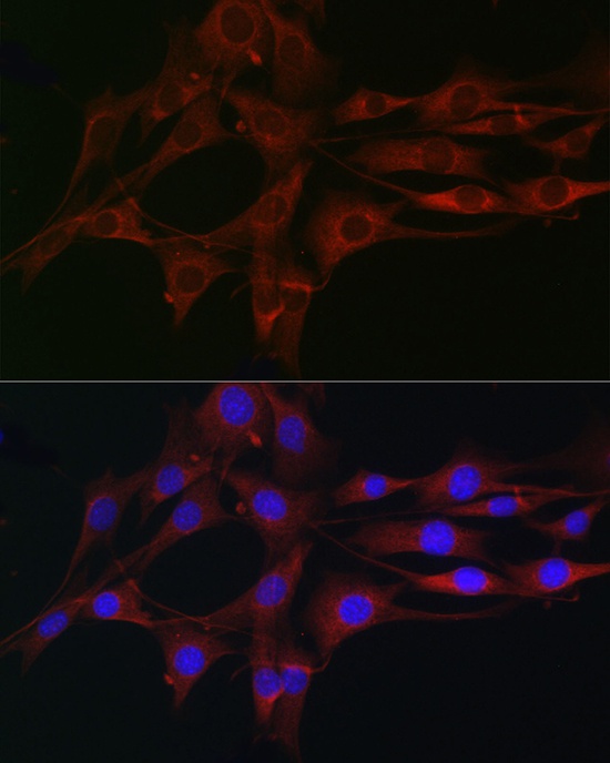
Immunofluorescence analysis of NIH/3T3 cells using CSK Rabbit pAb (CAB16824) at dilution of 1:100 (40x lens). Secondary antibody: Cy3-conjugated Goat anti-Rabbit IgG (H+L) (CABS007) at 1:500 dilution. Blue: DAPI for nuclear staining.