
Dendritic Cells - Markers, Activation & Subtypes
Dendritic Cells: Markers, Activation and Subtypes
Dendritic cells are the professional antigen-presenting cells that decide whether an immune response happens at all. They are also awkward to identify, because no single marker is exclusive to them — they are defined by a combination of positive markers and the absence of other lineages. This guide covers the markers that actually work, how activation and cross-presentation function, and the real subsets, including which cells share the name without sharing the lineage.
Browse DC marker antibodies →Key takeaways
- Dendritic cells are the only antigen-presenting cells that efficiently prime naive T cells, which makes them the initiators of adaptive immunity rather than just participants in it.
- No marker is exclusive to DCs. They are identified as CD11c-positive and HLA-DR-positive while lacking the lineage markers CD3, CD14, CD19 and CD56 — so CD14 and CD19 are exclusion markers, not DC markers.
- MHC class I presents peptides from cytosolic proteins to CD8 T cells; MHC class II presents peptides from internalised material to CD4 T cells. DCs additionally perform cross-presentation, routing external antigen onto MHC class I.
- The three principal human subsets are cDC1 (CD141-positive), cDC2 (CD1c-positive) and plasmacytoid DCs (CD123-positive).
- Plasmacytoid DCs are defined by massive type I interferon production — IFN-alpha and IFN-beta — not by interferon gamma, which comes mainly from T and NK cells.
- Follicular dendritic cells are not dendritic cells: they are stromal in origin and share only the name and the branched morphology.
- Maturation is the switch that matters therapeutically — immature DCs presenting self antigen induce tolerance, while matured DCs presenting the same antigen induce immunity.
Contents
- What is a dendritic cell?
- Function and antigen presentation
- Dendritic cell markers
- Activation and maturation
- Cytokines produced by DCs
- Dendritic cell subtypes
- Cells called dendritic that are not DCs
- Tolerogenic dendritic cells
- Dendritic cells and cancer
- References
- Choosing marker antibodies
- Frequently asked questions
Reagents for dendritic cell work
Because DCs are identified by marker combinations rather than any single antigen, the panel below covers the core gate, the subset-defining markers, and the functional readouts.
![APC Anti-Human CD11c Antibody [BU15]](https://cdn11.bigcommerce.com/s-h68l9z2lnx/products/20320/images/607360/apc-anti-human-cd11c-antibody-bu15-agel0448__84344.1707500940.386.513.jpg?c=2)
APC Anti-Human CD11c Antibody [BU15]
The principal conventional DC marker — used with HLA-DR to define the cDC gate.
View product →![Purified Anti-Human HLA-DR Antibody [L243]](https://cdn11.bigcommerce.com/s-h68l9z2lnx/products/20286/images/607088/anti-human-hla-dr-antibody-l243-agel0413__39967.1707500096.386.513.jpg?c=2)
Purified Anti-Human HLA-DR Antibody [L243]
MHC class II, expressed highly on mature DCs; the second half of the core gate.
View product →![Anti-CD1c [ZN46] Monoclonal Antibody](https://cdn11.bigcommerce.com/s-h68l9z2lnx/products/277485/images/679274/anti-cd1c-zn46-monoclonal-antibody-agmb06204__78345.1773038418.386.513.jpg?c=2)
Anti-CD1c [ZN46] Monoclonal Antibody
CD1c, also called BDCA-1, identifies the cDC2 subset — the most abundant conventional DC in blood.
View product →![GenieFluor 488 Anti-Human CD123 Antibody [6H6]](https://cdn11.bigcommerce.com/s-h68l9z2lnx/products/20317/images/606269/geniefluor-488-anti-human-cd123-antibody-6h6-agel0445__18610.1707497475.386.513.jpg?c=2)
GenieFluor 488 Anti-Human CD123 Antibody [6H6]
The IL-3 receptor alpha chain, expressed highly on plasmacytoid DCs.
View product →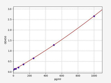
Human FLT3L ELISA Kit
FLT3 ligand is the essential growth factor for DC development; measuring it tracks differentiation capacity.
View product →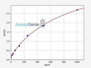
Human IL-12 p70 ELISA Kit
IL-12 is the signature cDC cytokine driving Th1 polarisation — the functional test of DC activation.
View product →What is a dendritic cell?
Dendritic cells are professional antigen-presenting cells that bridge innate and adaptive immunity. Like macrophages, they use pattern recognition receptors to detect pathogen-associated molecular patterns and internalise what they find. Unlike macrophages, their primary purpose is not to destroy the material but to process it and present it to T cells.
Their defining property is worth stating precisely: DCs are the only cells that efficiently prime naive T cells. Macrophages and B cells present antigen, but to T cells that have already been activated. Because a naive T cell requires a DC to be activated at all, DCs decide whether an adaptive response begins — which is why they are the target of therapeutic vaccines.
They are found in essentially every tissue, concentrated at barrier surfaces where pathogens enter, and they continuously sample their surroundings whether or not infection is present.
Function and antigen presentation
The DC life cycle has a clear direction: sample in the periphery, mature on detecting danger, migrate to a lymph node, present to T cells.
The two MHC routes
Antigen presentation is often described loosely, so it is worth being exact. MHC molecules do not recognise pathogens — they display short peptides, and which class a peptide reaches depends on where it came from:
| Pathway | Peptide source | Presented to |
|---|---|---|
| MHC class I | Proteins degraded in the cytosol, typically endogenous or from a virus replicating inside the cell | CD8-positive cytotoxic T cells |
| MHC class II | Material internalised from outside the cell and degraded in endosomes | CD4-positive helper T cells |
| Cross-presentation | Externally acquired antigen diverted into the MHC class I route | CD8-positive cytotoxic T cells |
Cross-presentation is the DC-specific capability, and its absence from most accounts is a real omission. It allows a DC that has never itself been infected to prime cytotoxic T cells against a virus or a tumour — by taking up material from a dying infected or malignant cell and routing it onto MHC class I. The cDC1 subset is the specialist at this, which is why cDC1 abundance predicts response to checkpoint immunotherapy.
The three signals
Presenting peptide alone does not activate a T cell. Three signals are needed: the peptide-MHC complex itself; costimulation through CD80 and CD86 engaging CD28; and cytokines that direct the resulting T cell’s phenotype. A DC delivering signal one without signals two and three induces tolerance rather than immunity — which is the basis of both self-tolerance and tolerogenic DC therapy.
Dendritic cell markers
This is where most identification errors occur, because no marker is exclusive to dendritic cells. They are defined by a combination of what they express and what they lack.
The lineage-negative requirement
DCs are identified as lineage-negative: they do not express CD3 (T cells), CD14 (monocytes), CD19 or CD20 (B cells), CD56 (NK cells) or CD66b (granulocytes). Those markers are therefore exclusion markers — a dump channel used to remove everything that is not a DC. Listing CD14 or CD19 among DC markers inverts their purpose, and is a common and consequential mistake: a gate built that way selects monocytes and B cells rather than DCs.
The positive markers
| Population | Human markers | Mouse markers | Note |
|---|---|---|---|
| Conventional DCs (core gate) | CD11c-positive, HLA-DR-positive, lineage-negative | CD11c-positive, MHC class II-positive | The starting gate for all cDC work |
| cDC1 | CD141 (BDCA-3), CLEC9A, XCR1 | CD8-alpha (lymphoid tissue), CD103 (peripheral), XCR1 | Specialists in cross-presentation; CD8-alpha is a mouse marker, not human |
| cDC2 | CD1c (BDCA-1), CD11b, CD172a | CD11b, CD172a | The most abundant conventional DC in human blood |
| Plasmacytoid DCs | CD123, CD303 (BDCA-2), CD304 (BDCA-4), CD45RA | B220, Ly-6C, Siglec-H, CD11c-low | Low MHC class II and low CD11c; TLR7 and TLR9 expressing |
| Maturation markers | CD80, CD86, CD83, CD40, high HLA-DR, CCR7 | CD80, CD86, CD40, MHC class II-high | Present on matured DCs regardless of subset |
Two nomenclature points that cause confusion. The BDCA series is human nomenclature and does not apply to mouse cells, so a mouse panel built on BDCA markers will not work. And CD8-alpha marks mouse cDC1, not human — the human equivalent is CD141/BDCA-3. Mixing the two species’ marker sets is the second commonest error after misusing the lineage markers.
Activation and maturation
DCs exist in two functional states, and the transition between them is the single most important event in DC biology.
Immature DCs are highly endocytic but poor presenters. They express low levels of MHC class II and costimulatory molecules, and they sample antigen continuously. Presenting self antigen in this state induces tolerance.
Maturation is triggered by pattern recognition receptor engagement — TLRs detecting microbial products, or damage signals from injured tissue. On maturation the DC reduces endocytosis, upregulates MHC class II, CD80, CD86, CD83 and CD40, and switches on CCR7 so that it migrates toward the CCL19 and CCL21 gradient leading to draining lymph nodes.
That coupling matters: the same signal that tells a DC an antigen is dangerous also gives it the means to reach T cells and the costimulation needed to activate them. Antigen encountered without danger signals never gets that treatment, which is how the system distinguishes threat from self.
Growth factors govern the supply of DCs rather than their activation. FLT3 ligand is essential for development of both conventional and plasmacytoid DCs, and GM-CSF with IL-4 is the standard combination for differentiating monocyte-derived DCs in vitro — the workhorse of most DC laboratory work, though monocyte-derived DCs are not equivalent to the DCs found in tissue.
Cytokines produced by DCs
| Cytokine | Source | Effect |
|---|---|---|
| IL-12 | Conventional DCs, especially cDC1 | Drives Th1 differentiation and activates NK cells; the signature cDC cytokine |
| Type I interferon (IFN-alpha, IFN-beta) | Plasmacytoid DCs | Produced in very large amounts on nucleic acid sensing; the defining pDC function |
| IL-23 | Conventional DCs | Sustains Th17 responses; central to psoriasis and inflammatory bowel disease |
| IL-6 | Conventional DCs | Contributes to Th17 differentiation and general inflammation |
| TNF-alpha | Conventional DCs | Amplifies local inflammation and promotes DC maturation |
| IL-10 and TGF-beta | Tolerogenic and regulatory DCs | Suppress responses and promote regulatory T cell induction |
One correction worth making explicitly: interferon gamma is not a dendritic cell cytokine in any meaningful sense. It is produced principally by T cells and NK cells, and it acts on DCs rather than being made by them. The interferon that defines DC biology is type I — IFN-alpha and IFN-beta from plasmacytoid DCs. The two are frequently conflated because both are called interferon.
Dendritic cell subtypes
Human dendritic cells divide into three well-defined populations, plus a monocyte-derived population generated in vitro.
Conventional dendritic cells
cDCs are the classical antigen-presenting DCs, expressing high MHC class I and class II. They split into two lineages. cDC1 cells are relatively rare, identified by CD141, CLEC9A and XCR1, and specialise in cross-presentation to CD8 T cells — which makes them disproportionately important in antitumour immunity. cDC2 cells are more abundant, identified by CD1c, and are more versatile, driving Th1, Th2 and Th17 responses depending on context.
Plasmacytoid dendritic cells
pDCs circulate in blood and populate lymphoid tissue. They express low CD11c and low MHC class II, and are weak antigen presenters by comparison with cDCs. Their function is different: expressing TLR7 and TLR9, they sense viral RNA and DNA and respond by secreting type I interferon in quantities far exceeding any other cell type. They are effectively a dedicated antiviral alarm.
Monocyte-derived dendritic cells
Differentiated from blood monocytes with GM-CSF and IL-4, moDCs are the practical workhorse of DC research and of most clinical DC vaccine protocols, simply because large numbers can be generated from a blood draw. They are genuinely DC-like but transcriptionally distinct from tissue cDCs, so findings should not be assumed to transfer.
Langerhans cells
Resident in the epidermis and often described as epidermal dendritic cells, Langerhans cells are identified by CD1a and Langerin (CD207). They are unusual in origin — they arise from embryonic precursors and self-renew locally rather than being continuously replaced from blood, which makes them closer to tissue-resident macrophages developmentally while functioning as antigen-presenting cells. They contribute to contact hypersensitivity and are implicated in psoriasis and atopic dermatitis.
Cells called dendritic that are not DCs
Two populations carry the name without belonging to the lineage, and conflating them with real DCs causes genuine confusion.
Follicular dendritic cells are not dendritic cells. They are of stromal, mesenchymal origin — not haematopoietic — and share only the branched morphology that gave both cell types their name. Their function is also different: rather than processing antigen and presenting peptide on MHC, FDCs retain intact antigen in immune complexes on their surface using complement receptors CD21 and CD35 and Fc receptors. That display allows B cells in germinal centres to compete for antigen, which is how affinity maturation selects for higher-affinity antibody. They support B cell selection; they do not prime T cells.
Interdigitating dendritic cells is older terminology for the interdigitating reticulum cells of lymph node T cell zones. In modern usage these are largely regarded as conventional DCs described by their histological location. They present antigen to T cells and do not produce antibodies — antibody production is exclusive to B cells and plasma cells, and any statement that DCs make antibodies is incorrect.
Tolerogenic dendritic cells
Tolerogenic DCs actively suppress rather than initiate immune responses, and they are a functional state rather than a separate lineage. A DC presenting antigen without adequate costimulation, or under the influence of IL-10, TGF-beta, vitamin D or corticosteroids, induces tolerance instead of immunity.
Their mechanisms include low costimulatory molecule expression, secretion of IL-10 and TGF-beta, expression of inhibitory ligands such as PD-L1, and induction of regulatory T cells. Both conventional and plasmacytoid DCs can adopt a tolerogenic phenotype — pDCs are notable for IL-10 production and for inducing Tregs.
A clarification on terminology: NK cells are not a type of dendritic cell, tolerogenic or otherwise. They are cytotoxic innate lymphocytes of a separate lineage. Any classification listing tolerogenic NK cells among DC subtypes is a category error, though NK cells and DCs do communicate bidirectionally.
Clinically, tolerogenic DCs are being explored for autoimmune disease and transplant tolerance — the inverse application of the DC vaccines used in cancer.
Dendritic cells and cancer
DCs matter in oncology in three distinct ways.
As the initiators of antitumour immunity
Cross-presentation by cDC1 cells is how cytotoxic T cell responses against tumour antigens begin. Tumours that exclude or disable cDC1 cells are correspondingly harder to treat, and cDC1 abundance within a tumour is associated with response to checkpoint inhibition — because checkpoint blockade releases T cells that a DC must first have primed.
As a therapeutic platform
DC vaccines load patient-derived DCs with tumour antigen ex vivo and reinfuse them. The approach is mechanistically sound and clinically approved in prostate cancer, though results across tumour types have been mixed — attributed variously to the use of monocyte-derived rather than cDC1 cells, insufficient maturation, and the immunosuppressive tumour microenvironment blunting whatever response is generated.
As the origin of malignancy
| Condition | Origin | Notes |
|---|---|---|
| Blastic plasmacytoid dendritic cell neoplasm (BPDCN) | Plasmacytoid DC precursors | Rare and aggressive; median age at diagnosis around 65–70, male predominance. Usually presents with skin lesions before marrow involvement. Arises from somatic mutations — it is not an inherited cancer |
| Follicular dendritic cell sarcoma | Follicular dendritic cells (stromal, not haematopoietic) | Very rare; frequently head and neck. Treated primarily with surgery, sometimes with radiotherapy. Generally better prognosis than BPDCN, varying with size and site |
| Langerhans cell histiocytosis | Langerhans cell lineage | Frequently driven by BRAF V600E mutation, which has made BRAF inhibition a treatment option |
Diagnosis and management of these conditions are specialist matters; this section describes the cell biology and is not clinical guidance.
References
- Mellman I. Dendritic cells: master regulators of the immune response. Cancer Immunology Research, 2013;1(3):145–149.
- Vyas JM. The dendritic cell: the general of the army. Virulence, 2012;3(7):601–602.
- Hellman P, Eriksson H. Early activation markers of human peripheral dendritic cells. Human Immunology, 2007;68(5):324–333.
- Nunez R. Assessment of surface markers and functionality of dendritic cells. Current Protocols in Cytometry, 2001; Unit 9.17.
- Ueno H, Klechevsky E, Morita R, et al. Dendritic cell subsets in health and disease. Immunological Reviews, 2007;219:118–142.
Choosing marker antibodies
Conjugated antibodies against CD11c, HLA-DR, CD1c and CD123 for gating conventional and plasmacytoid DCs, plus FLT3-L and IL-12 assays for development and functional readouts.
Browse DC marker antibodies →Frequently asked questions
What markers identify dendritic cells?
A combination. DCs are CD11c-positive and HLA-DR-positive while lacking the lineage markers CD3, CD14, CD19, CD56 and CD66b. Subsets are then resolved with CD141 for cDC1, CD1c for cDC2 and CD123 for plasmacytoid DCs. No single marker is exclusive to DCs.
Are CD14 and CD19 dendritic cell markers?
No — the opposite. CD14 marks monocytes and CD19 marks B cells, and both are used as exclusion markers to remove those populations when gating DCs. Treating them as DC markers produces a gate that selects monocytes and B cells instead.
What is cross-presentation?
The routing of externally acquired antigen onto MHC class I, allowing a DC to prime cytotoxic CD8 T cells against a pathogen or tumour it was never itself infected by. The cDC1 subset specialises in it, which is why cDC1 abundance associates with checkpoint inhibitor response.
Are follicular dendritic cells dendritic cells?
No. They are stromal, mesenchymal in origin rather than haematopoietic, and share only the branched morphology and the name. They retain intact antigen in immune complexes for B cell selection in germinal centres rather than presenting peptide to T cells.
Do dendritic cells produce interferon gamma?
Not meaningfully. Interferon gamma comes chiefly from T cells and NK cells and acts on DCs. The interferon that defines DC biology is type I — IFN-alpha and IFN-beta produced in very large amounts by plasmacytoid DCs.
What is the difference between immature and mature DCs?
Immature DCs sample antigen avidly but express little MHC class II or costimulation, and presenting self antigen in that state induces tolerance. Maturation, triggered by pattern recognition receptors, upregulates MHC and costimulation and switches on CCR7 for migration to lymph nodes.
Why are monocyte-derived DCs used if they are not real tissue DCs?
Because they can be generated in large numbers from a routine blood draw using GM-CSF and IL-4, which tissue cDCs cannot. They are genuinely DC-like and underpin most clinical DC vaccine protocols, but they are transcriptionally distinct from tissue cDCs, so findings do not automatically transfer.
Pragna Krishnapur completed her bachelor degree in Biotechnology Engineering at Visvesvaraya Technological University before completing her masters in Biotechnology at University College Dublin.
Recent Posts
-
Suspected Plague Death at a Siberian Anti-Plague Institute: What Researchers Should Know
Infectious Disease · News AnalysisSuspected Plague Death at a Siberian Anti-Plague Institute: What …6th Oct 2026 -
LPS ELISA Troubleshooting: The Importance of Sample Nature
Quick answer A researcher measuring circulating lipopolysaccharide (LPS) in human serum …25th Sep 2026 -

Sekundärantikörper wählen: Die Regel, die im Labor wirklich zählt
Sekundärantikörper wählen: Die Regel, die im Labor wirklich zählt Wählen Sie einen Sekundärantikörp …24th Sep 2026