The FGF1 Monoclonal Antibody (CAB5900) is a high-quality antibody developed for reliable detection and analysis of target proteins. This antibody, developed using rabbit monoclonal technology, shows high specificity and sensitivity for detecting FGF1 in human samples.With its validated performance in Western blot applications, this antibody enables precise detection and quantification of FGF1 in various cell types and tissues. Its ability to specifically bind to the FGF1 protein opens up opportunities for detailed analysis and research in fields such as developmental biology, cancer biology, and regenerative medicine.
This antibody is validated for use in WB, IF/ICC, ELISA applications and has demonstrated reactivity against Human, Mouse, Rat samples.
Product Name:
FGF1 Monoclonal Antibody
SKU:
CAB5900
Size:
20μL, 100μL
Reactivity:
Human, Mouse, Rat
Clone Number:
ARC1414
Conjugate:
Unconjugated
Immunogen:
Synthetic peptide. This information is considered to be commercially sensitive.
The protein encoded by this gene is a member of the fibroblast growth factor (FGF) family. FGF family members possess broad mitogenic and cell survival activities, and are involved in a variety of biological processes, including embryonic development, cell growth, morphogenesis, tissue repair, tumor growth and invasion. This protein functions as a modifier of endothelial cell migration and proliferation, as well as an angiogenic factor. It acts as a mitogen for a variety of mesoderm- and neuroectoderm-derived cells in vitro, thus is thought to be involved in organogenesis. Multiple alternatively spliced variants encoding different isoforms have been described.
Purification Method
Affinity purification
Gene ID
2246
RRID
AB_2863520
Buffer Information
Store at -20℃. Avoid freeze / thaw cycles. Buffer: PBS containing 50% glycerol and 0.05% BSA, preserved with proclin300 or sodium azide, pH 7.3.
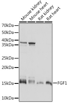
Western blot analysis of various lysates using FGF1 Rabbit mAb (CAB5900) at 1:1000 dilution. Secondary antibody: HRP-conjugated Goat anti-Rabbit IgG (H+L) (CABS014) at 1:10000 dilution. Lysates/proteins: 25μg per lane. Blocking buffer: 3% nonfat dry milk in TBST. Detection: ECL Basic Kit (AbGn00020). Exposure time: 10s.
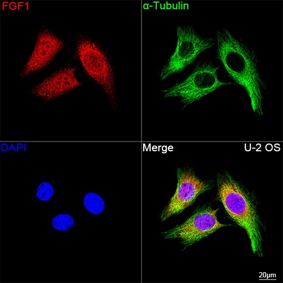
Confocal imaging of U-2 OS cells using FGF1 Rabbit mAb (CAB5900, dilution 1:200) followed by a further incubation with Cy3 Goat Anti-Rabbit IgG (H+L) (CABS007, dilution 1:500) (Red). The cells were counterstained with α-Tubulin Mouse mAb (AC012, dilution 1:400) followed by incubation with ABflo® 488-conjugated Goat Anti-Mouse IgG (H+L) Ab (CABS076, dilution 1:500) (Green). DAPI was used for nuclear staining (Blue). Objective: 100x.
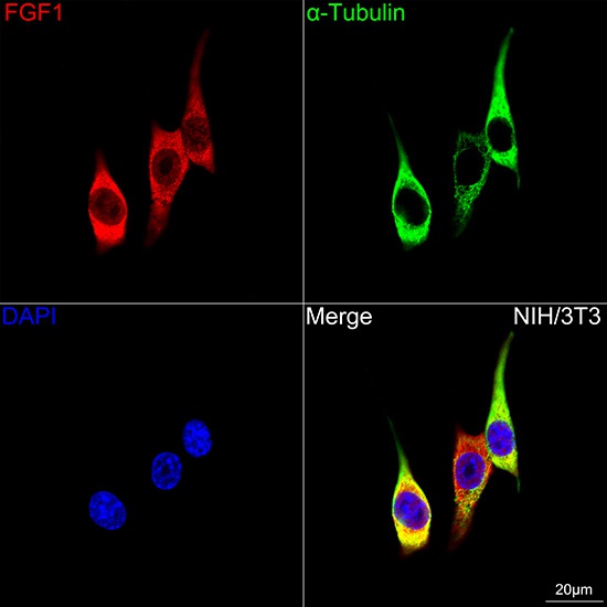
Confocal imaging of NIH/3T3 cells using FGF1 Rabbit mAb (CAB5900, dilution 1:200) followed by a further incubation with Cy3 Goat Anti-Rabbit IgG (H+L) (CABS007,dilution 1:500) (Red).The cells were counterstained with α-Tubulin Mouse mAb (AC012, dilution 1:400) followed by incubation with ABflo® 488-conjugated Goat Anti-Mouse IgG (H+L) Ab (CABS076, dilution 1:500) (Green). DAPI was used for nuclear staining (Blue). Objective: 100x.


![Anti-FGF1 [R05-6A4] Monoclonal Antibody (AGMB02122)](https://cdn11.bigcommerce.com/s-h68l9z2lnx/images/stencil/590x590/products/273411/677106/anti-fgf1-r05-6a4-monoclonal-antibody-agmb02122__84970.1773031580.jpg?c=2 "Anti-FGF1 [R05-6A4] Monoclonal Antibody (AGMB02122)")
![Anti-FGF1 Rat [16C6] Monoclonal Antibody (AGMB04655)](https://cdn11.bigcommerce.com/s-h68l9z2lnx/images/stencil/590x590/products/275940/679788/anti-fgf1-rat-16c6-monoclonal-antibody-agmb04655__90090.1773039996.jpg?c=2 "Anti-FGF1 Rat [16C6] Monoclonal Antibody (AGMB04655)")
![Anti-FGF1 [R02-9V5] Monoclonal Antibody (AGMB02948)](https://cdn11.bigcommerce.com/s-h68l9z2lnx/images/stencil/590x590/products/274237/681019/anti-fgf1-r02-9v5-monoclonal-antibody-agmb02948__86880.1773043936.jpg?c=2 "Anti-FGF1 [R02-9V5] Monoclonal Antibody (AGMB02948)")

