The [KO Validated] FUS Antibody (CAB5921) is a high-quality antibody developed for reliable detection and analysis of target proteins. This antibody, produced in rabbits, has been rigorously tested and validated for knockout validation, ensuring reliable results in various experimental settings.FUS, also known as fused in sarcoma, is a multifunctional RNA-binding protein involved in mRNA splicing, transcriptional regulation, and DNA repair. Dysregulation of FUS has been linked to diseases like amyotrophic lateral sclerosis (ALS) and frontotemporal dementia (FTD), making it a promising target for therapeutic intervention.
This antibody is validated for use in WB, IF/ICC, IP, ELISA applications and has demonstrated reactivity against Human, Mouse, Rat samples.
Product Name:
[KO Validated] FUS Antibody
SKU:
CAB5921
Size:
20μL, 100μL
Reactivity:
Human, Mouse, Rat
Conjugate:
Unconjugated
Immunogen:
Recombinant protein (or fragment).This information is considered to be commercially sensitive.
0.5μg-4μg antibody for 200μg-400μg extracts of whole cells
ELISA
Recommended starting concentration is 1 μg/mL. Please optimize the concentration based on your specific assay requirements.
Synonyms:
TLS, ALS6, ETM4, FUS1, POMP75, altFUS, HNRNPP2, US
Positive Sample:
U-937, Jurkat
Cellular Localization:
Nucleus.
Calculated MW:
53kDa
Observed MW:
70kDa
This gene encodes a multifunctional protein component of the heterogeneous nuclear ribonucleoprotein (hnRNP) complex. The hnRNP complex is involved in pre-mRNA splicing and the export of fully processed mRNA to the cytoplasm. This protein belongs to the FET family of RNA-binding proteins which have been implicated in cellular processes that include regulation of gene expression, maintenance of genomic integrity and mRNA/microRNA processing. Alternative splicing results in multiple transcript variants. Defects in this gene result in amyotrophic lateral sclerosis type 6.
Purification Method
Affinity purification
Gene ID
2521
RRID
AB_2863521
Buffer Information
Store at -20℃. Avoid freeze / thaw cycles. Buffer: PBS containing 50% glycerol, preserved with proclin300 or sodium azide, pH 7.3.
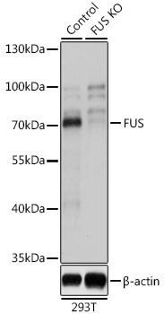
Western blot analysis of lysates from wild type (WT) and FUS knockout (KO) 293T cells, using [KO Validated] FUS Rabbit pAb (CAB5921) at 1:3000 dilution. Secondary antibody: HRP-conjugated Goat anti-Rabbit IgG (H+L) (CABS014) at 1:10000 dilution. Lysates/proteins: 25μg per lane. Blocking buffer: 3% nonfat dry milk in TBST. Detection: ECL Basic Kit (AbGn00020). Exposure time: 1s.
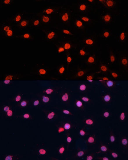
Immunofluorescence analysis of C6 cells using [KO Validated] FUS Rabbit pAb (CAB5921) at dilution of 1:100. Secondary antibody: Cy3-conjugated Goat anti-Rabbit IgG (H+L) (CABS007) at 1:500 dilution. Blue: DAPI for nuclear staining.
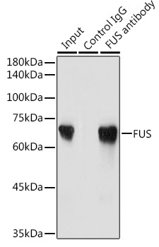
Immunoprecipitation analysis of 300 μg extracts of Jurkat cells using 3 μg FUS antibody (CAB5921). Western blot was performed from the immunoprecipitate using FUS antibody (CAB5921) at a dilution of 1:2000.