Description
Bradford Protein Assay (BA0168) (BA0168)
The Bradford Protein Assay (SKU: BA0168) provides a simple, direct and automation-ready procedure for measuring total protein concentration. Proteins are polypeptides made up of amino acids that play key roles in all aspects of biology, and protein quantitation is a very common practice in the life sciences. This kit is based on an improved Coomassie Blue G method, in which the dye forms a blue complex specifically with protein; the intensity of colour, measured at 595 nm, is directly proportional to the protein concentration in the sample. The optimised formulation substantially reduces interference by substances in raw samples and exhibits increased sensitivity towards peptides. The mix-and-read procedure uses 10 uL of sample and can be readily automated as a high-throughput assay for thousands of samples per day.
| Product Name: | Bradford Protein Assay (BA0168) |
| SKU: | BA0168 |
| Detection Method: | Colorimetric (Bradford) |
| Detection Range: | 0.06 - 1.0 mg/mL protein |
| Sample Type: | Protein samples including tissue and other biological samples |
| Species Reactivity: | All |
| Assay Time: | Immediate (mix-and-read) |
| Kit Size: | 500 Assays |
| Equipment Required: | Microplate reader |
| Storage: | Store the reagent at 4 C and the standard at -20 C. |
| Shelf Life: | 12 months after receipt |
| Shipping: | Room Temperature |
A colorimetric assay for the quantitative determination of total protein based on an improved Coomassie Blue G (Bradford) method. The dye forms a blue complex with protein measured at 595 nm, with intensity proportional to protein concentration. The mix-and-read format uses small sample volumes and is readily automated for high-throughput use.
- Sensitive and accurate, using 10 uL of sample, with a detection range of 0.06 - 1.0 mg/mL protein
- Simple mix-and-read format with a single working reagent
- Low interference from common buffer components
- Versatile, with 96-well plate and cuvette formats
- Direct determination of total protein concentration
Note: The below protocol is a sample protocol. Protocols are specific to each batch/lot. For the correct instructions please follow the protocol included in your kit.
| Step | Procedure |
| 1 | Prepare the working reagent by adding 1 volume of the 5x Reagent to 5 volumes of distilled water and bringing it to room temperature before use. |
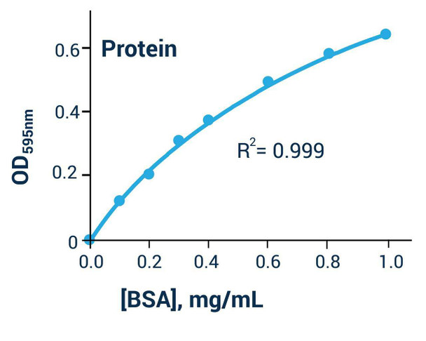
| 2 | Dilute the standard to 1.0, 0.8, 0.6, 0.4, 0.3, 0.2, 0.1 and 0 mg/mL BSA. Transfer 10 uL of each diluted standard and 10 uL of each diluted sample in duplicate into wells of a clear-bottom 96-well plate. |
| 3 | Add 200 uL working reagent to each well and tap lightly to mix. |
| 4 | Measure optical density at 570-630 nm (peak 595 nm). |
| 5 | For the cuvette format, transfer 50 uL of standards and samples to cuvettes, add 1000 uL working reagent and measure optical density at 570-630 nm (peak 595 nm). |
Subtract the water blank from the standard values and plot the optical density against protein concentration. Use the standard curve to determine the sample protein concentration. If the concentration exceeds 1 mg/mL, dilute the sample in distilled water and use readings within the calibration range.
| Component | Quantity | Storage |
| Reagent (5x concentrate) | 20 mL | 4 C |
| Protein Standard (1.0 mg/mL BSA) | 1 mL | -20 C |