The IGF2BP3 Antibody (CAB6099) is a high-quality antibody developed for reliable detection and analysis of target proteins. This antibody, generated in rabbits, exhibits high reactivity with human samples and has been validated for use in Western blot applications.IGF2BP3, also known as IMP3, is a member of the insulin-like growth factor-2 mRNA-binding protein family and plays a critical role in post-transcriptional regulation of gene expression. It is overexpressed in various cancers, including pancreatic, lung, and ovarian cancer, and its expression has been linked to poor prognosis in cancer patients.
This antibody is validated for use in WB, IHC-P, IF/ICC, IP, ELISA applications and has demonstrated reactivity against Human, Mouse, Rat samples.
Product Name:
IGF2BP3 Antibody
SKU:
CAB6099
Size:
20μL, 100μL
Reactivity:
Human, Mouse, Rat
Conjugate:
Unconjugated
Immunogen:
Recombinant protein (or fragment).This information is considered to be commercially sensitive.
0.5μg-4μg antibody for 200μg-400μg extracts of whole cells
ELISA
Recommended starting concentration is 1 μg/mL. Please optimize the concentration based on your specific assay requirements.
Synonyms:
KOC, CT98, IMP3, KOC1, IMP-3, VICKZ3, IGF2BP3
Positive Sample:
HeLa, T-47D, Rat testis
Cellular Localization:
Cytoplasm, Nucleus.
Calculated MW:
64kDa
Observed MW:
64kDa
The protein encoded by this gene is primarily found in the nucleolus, where it can bind to the 5' UTR of the insulin-like growth factor II leader 3 mRNA and may repress translation of insulin-like growth factor II during late development. The encoded protein contains several KH domains, which are important in RNA binding and are known to be involved in RNA synthesis and metabolism. A pseudogene exists on chromosome 7, and there are putative pseudogenes on other chromosomes.
Purification Method
Affinity purification
Gene ID
10643
RRID
AB_2766745
Buffer Information
Store at -20℃. Avoid freeze / thaw cycles. Buffer: PBS containing 50% glycerol, preserved with proclin300 or sodium azide, pH 7.3.
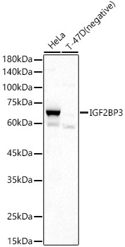
Western blot analysis of various lysates, using IGF2BP3 Rabbit pAb (CAB6099) at 1:500 dilution. Secondary antibody: HRP-conjugated Goat anti-Rabbit IgG (H+L) (CABS014) at 1:10000 dilution. Lysates/proteins: 25μg per lane. Blocking buffer: 3% nonfat dry milk in TBST. Detection: ECL Basic Kit (AbGn00020). Exposure time: 15s.
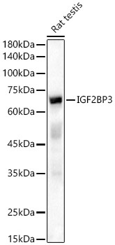
Western blot analysis of lysates from Rat testis, using IGF2BP3 Rabbit pAb (CAB6099) at 1:500 dilution. Secondary antibody: HRP-conjugated Goat anti-Rabbit IgG (H+L) (CABS014) at 1:10000 dilution. Lysates/proteins: 25μg per lane. Blocking buffer: 3% nonfat dry milk in TBST. Detection: ECL Basic Kit (AbGn00020). Exposure time: 15s.
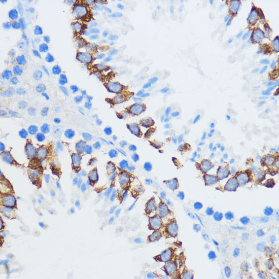
Immunohistochemistry analysis of paraffin-embedded Mouse testis using IGF2BP3 Rabbit pAb (CAB6099) at dilution of 1:100 (40x lens). Microwave antigen retrieval performed with 0.01M PBS Buffer (pH 7.2) prior to IHC staining.
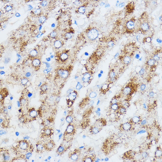
Immunohistochemistry analysis of paraffin-embedded Human liver using IGF2BP3 Rabbit pAb (CAB6099) at dilution of 1:100 (40x lens). Microwave antigen retrieval performed with 0.01M PBS Buffer (pH 7.2) prior to IHC staining.
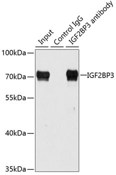
Immunoprecipitation analysis of 100 μg extracts of HepG2 cells using 3 μg IGF2BP3 antibody (CAB6099). Western blot was performed from the immunoprecipitate using IGF2BP3 antibody (CAB6099) at a dilution of 1:1000.