The [KO Validated] RCN2 Antibody (CAB19943) is a high-quality antibody developed for reliable detection and analysis of target proteins. This rabbit-derived antibody is highly specific for detecting RCN2 in human samples and has been validated for use in Western blot applications. By targeting the RCN2 protein, researchers can investigate its role in cellular processes related to protein quality control and calcium signaling.RCN2, also known as the reticulocalbin-2 protein, is essential for maintaining proper intracellular calcium levels and ensuring correct protein folding in the ER. Dysregulation of RCN2 has been linked to various diseases, including cancer and neurodegenerative disorders.
This antibody is validated for use in WB, ELISA applications and has demonstrated reactivity against Human, Mouse, Rat samples.
Product Name:
[KO Validated] RCN2 Antibody
SKU:
CAB19943
Size:
20μL, 100μL
Reactivity:
Human, Mouse, Rat
Conjugate:
Unconjugated
Immunogen:
Recombinant protein (or fragment).This information is considered to be commercially sensitive.
Recommended starting concentration is 1 μg/mL. Please optimize the concentration based on your specific assay requirements.
Synonyms:
E6BP, ERC55, ERC-55, TCBP49, N2
Positive Sample:
293T
Cellular Localization:
Endoplasmic Reticulum Lumen.
Calculated MW:
37kDa
Observed MW:
47kDa
The protein encoded by this gene is a calcium-binding protein located in the lumen of the ER. The protein contains six conserved regions with similarity to a high affinity Ca(+2)-binding motif, the EF-hand. This gene maps to the same region as type 4 Bardet-Biedl syndrome, suggesting a possible causative role for this gene in the disorder. Alternatively spliced transcript variants encoding different isoforms have been found for this gene.
Purification Method
Affinity purification
Gene ID
5955
RRID
AB_2862853
Buffer Information
Store at -20℃. Avoid freeze / thaw cycles. Buffer: PBS containing 50% glycerol, preserved with proclin300 or sodium azide, pH 7.3.
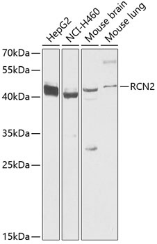
Western blot analysis of various lysates using RCN2 Rabbit pAb (CAB19943) at 1:1000 dilution. Secondary antibody: HRP-conjugated Goat anti-Rabbit IgG (H+L) (CABS014) at 1:10000 dilution. Lysates/proteins: 25μg per lane. Blocking buffer: 3% nonfat dry milk in TBST. Detection: ECL Basic Kit (AbGn00020). Exposure time: 10s.
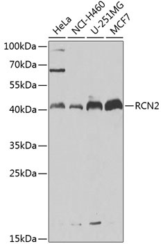
Western blot analysis of various lysates using RCN2 Rabbit pAb (CAB19943) at 1:1000 dilution. Secondary antibody: HRP-conjugated Goat anti-Rabbit IgG (H+L) (CABS014) at 1:10000 dilution. Lysates/proteins: 25μg per lane. Blocking buffer: 3% nonfat dry milk in TBST. Detection: ECL Basic Kit (AbGn00020). Exposure time: 30s.