The RHOT2 Antibody (CAB2597) is a high-quality antibody developed for reliable detection and analysis of target proteins. This antibody, generated in rabbits, exhibits high reactivity towards human samples and has been validated for Western blot applications. By binding specifically to Rhotekin 2, this antibody enables accurate detection and analysis of the protein in a variety of cell types, making it an essential tool for studies in cell biology and cancer research.Rhotekin 2 is known to play a crucial role in regulating cell migration, adhesion, and cytoskeletal organization, making it a key player in processes like tumor invasion and metastasis.
This antibody is validated for use in WB, IHC-P, IF/ICC, ELISA applications and has demonstrated reactivity against Human, Mouse, Rat samples.
Product Name:
RHOT2 Antibody
SKU:
CAB2597
Size:
20μL, 100μL
Reactivity:
Human, Mouse, Rat
Conjugate:
Unconjugated
Immunogen:
Recombinant protein (or fragment).This information is considered to be commercially sensitive.
Recommended starting concentration is 1 μg/mL. Please optimize the concentration based on your specific assay requirements.
Synonyms:
RASL, ARHT2, MIRO2, MIRO-2, C16orf39, RHOT2
Positive Sample:
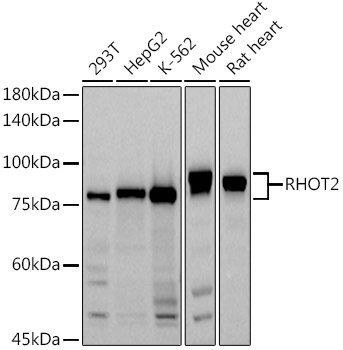
293T, HepG2, K-562, Mouse heart, Rat heart
Cellular Localization:
Mitochondrion Outer Membrane, Single-Pass Type Iv Membrane Protein.
Calculated MW:
68kDa
Observed MW:
80kDa
This gene encodes a member of the Rho family of GTPases. The encoded protein is localized to the outer mitochondrial membrane and plays a role in mitochondrial trafficking and fusion-fission dynamics.
Purification Method
Affinity purification
Gene ID
89941
RRID
AB_2764482
Buffer Information
Store at -20℃. Avoid freeze / thaw cycles. Buffer: PBS containing 50% glycerol, preserved with proclin300 or sodium azide, pH 7.3.
Western blot analysis of various lysates using (CAB2597) at 1:1000 dilution. Secondary antibody: HRP-conjugated Goat anti-Rabbit IgG (H+L) (CABS014) at 1:10000 dilution. Lysates/proteins: 25μg per lane. Blocking buffer: 3% nonfat dry milk in TBST. Detection: ECL Basic Kit (AbGn00020). Exposure time: 30s.
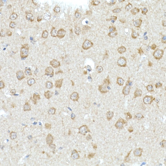
Immunohistochemistry analysis of paraffin-embedded Mouse brain using RHOT2 Rabbit pAb (CAB2597) at dilution of 1:50 (40x lens). High pressure antigen retrieval performed with 0.01M Citrate buffer (pH 6.0) prior to IHC staining.
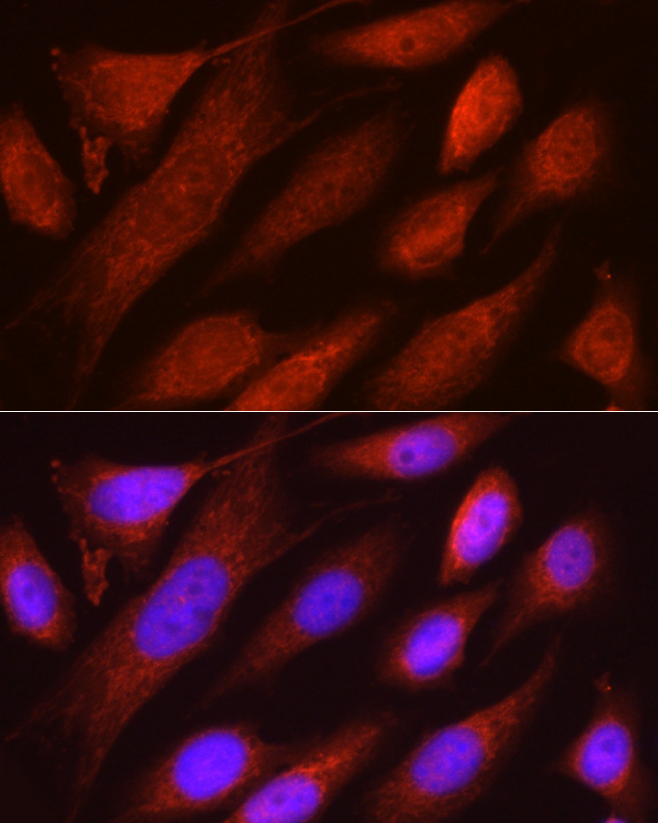
Immunofluorescence analysis of U2OS cells using RHOT2 Rabbit pAb (CAB2597) at dilution of 1:50 (40x lens). Secondary antibody: Cy3-conjugated Goat anti-Rabbit IgG (H+L) (CABS007) at 1:500 dilution. Blue: DAPI for nuclear staining.