The [KO Validated] HK1 Antibody (CAB1054) is a high-quality antibody developed for reliable detection and analysis of target proteins. This antibody, developed using rabbit immunization, exhibits high reactivity with human samples and has been specifically validated for use in Western blot applications.Hexokinase-1 plays a crucial role in catalyzing the first step of glucose metabolism, converting glucose to glucose-6-phosphate. Dysregulation of Hexokinase-1 activity has been linked to various diseases, including cancer and diabetes, making it an attractive target for therapeutic development and research.
This antibody is validated for use in WB, IHC-P, IF/ICC, IP, ELISA applications and has demonstrated reactivity against Human, Mouse, Rat samples.
Product Name:
[KO Validated] HK1 Antibody
SKU:
CAB1054
Size:
20μL, 100μL
Reactivity:
Human, Mouse, Rat
Conjugate:
Unconjugated
Immunogen:
Recombinant protein (or fragment).This information is considered to be commercially sensitive.
293T, MCF7, U-87MG, Mouse testis, Mouse brain, Mouse heart, Rat testis, Rat brain
Cellular Localization:
Mitochondrion Outer Membrane.
Calculated MW:
102kDa
Observed MW:
102kDa
Hexokinases phosphorylate glucose to produce glucose-6-phosphate, the first step in most glucose metabolism pathways. This gene encodes a ubiquitous form of hexokinase which localizes to the outer membrane of mitochondria. Mutations in this gene have been associated with hemolytic anemia due to hexokinase deficiency. Alternative splicing of this gene results in several transcript variants which encode different isoforms, some of which are tissue-specific.
Purification Method
Affinity purification
Gene ID
3098
RRID
AB_2758080
Buffer Information
Store at -20℃. Avoid freeze / thaw cycles. Buffer: PBS containing 50% glycerol, preserved with proclin300 or sodium azide, pH 7.3.
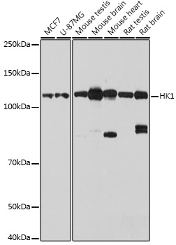
Western blot analysis of various lysates using [KO Validated] HK1 Rabbit pAb (CAB1054) at 1:1000 dilution. Secondary antibody: HRP-conjugated Goat anti-Rabbit IgG (H+L) (CABS014) at 1:10000 dilution. Lysates/proteins: 25μg per lane. Blocking buffer: 3% nonfat dry milk in TBST. Detection: ECL Basic Kit (AbGn00020). Exposure time: 10s.
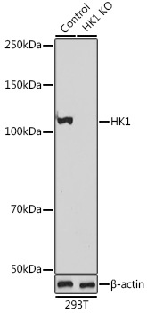
Western blot analysis of lysates from wild type (WT) and HK1 knockout (KO) 293T cells, using [KO Validated] HK1 Rabbit pAb (CAB1054) at 1:1000 dilution. Secondary antibody: HRP-conjugated Goat anti-Rabbit IgG (H+L) (CABS014) at 1:10000 dilution. Lysates/proteins: 25μg per lane. Blocking buffer: 3% nonfat dry milk in TBST. Detection: ECL Basic Kit (AbGn00020). Exposure time: 10s.
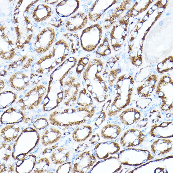
Immunohistochemistry analysis of paraffin-embedded Mouse kidney using HK1 Rabbit pAb (CAB1054) at dilution of 1:100 (40x lens). High pressure antigen retrieval performed with 0.01M Citrate buffer (pH 6.0) prior to IHC staining.
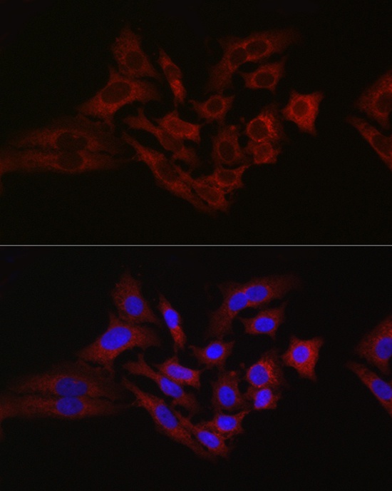
Immunofluorescence analysis of A-549 cells using [KO Validated] HK1 Rabbit pAb (CAB1054) at dilution of 1:100 (40x lens). Secondary antibody: Cy3-conjugated Goat anti-Rabbit IgG (H+L) (CABS007) at 1:500 dilution. Blue: DAPI for nuclear staining.
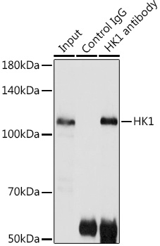
Immunoprecipitation analysis of 300 μg extracts of 293T cells using 3 μg HK1 antibody (CAB1054). Western blot was performed from the immunoprecipitate using HK1 antibody (CAB1054) at a dilution of 1:500.